The intestinal lamina propria contains many types of myeloid and lymphoid cells that maintain tolerance or carry out inflammatory responses. Click on these tabs for flow cytometry phenotyping suggestions and extended lists of secreted, cell surface, and intracellular markers. Each link will direct you to our product offerings for the study of these cells and molecules. For a broader representation of intestinal immunity, please view our poster Mucosal Immunity of the Intestine.
Lamina Propria Immune Cell Overview- Myeloid Cells in Homeostasis
- Myeloid Cells in Inflammation
- T Lineage Cells
- ILC in Homeostasis
- ILC in Inflammation
- Follicular Dendritic Cells
- B Lineage Cells
- Intraepithelial Lymphocytes
Phenotyping
Human
- CD11c+
- CX3CR1+
- CD3-
- CD14-
- CD19-
- MS4A1/CD20-
- NCAM-1/CD56-
Mouse
- CX3CR1+
- F4/80/EMR1+
- CD3-
- CD11c-
- CD14-
- CD19-
- MS4A1/CD20-
- NCAM-1/CD56-
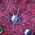
CD103+ Dendritic Cell
Additional Markers
- Secreted
- IL-10
- IL-22
- Retinoic Acid Agonists
- Retinoic Acid Antagonists
- TSLP
- Cell Surface
- CCR5
- CCR5 Antagonists
- CCR6
- CCR7
- CCR9
- CD25/IL-2 R alpha
- CD11c
- CD83
- CLEC9a
- Connexin 43/GJA1
- CXCR4
- DCIR/CLEC4a
- DC-SIGN/CD209
- F4/80/EMR1
- HLA-DR
- ICAM-1/CD54
- ICAM-1/CD54 Inhibitors
- IGSF4A/SynCAM1
- Integrin alpha 4
- Integrin alpha E/ CD103
- Integrin alpha M/ CD11b
- Integrin alpha M/ CD11b Activators
- Integrin alpha V beta 8
- Integrin beta 7
- Jagged 2
- Langerin/CD207
- MHC class II
(I-A/I-E) - OX40 Ligand/TNFSF4
- SIGNR1/CD209b
- SIRP alpha/CD172a
- Thrombomodulin/ BDCA-3
- TLR2
- TLR2 Inhibitors
- TLR4
- TLR4/MD-2 Complex
- TLR4 Inhibitors
- TLR5
- TLR7
- TLR7 Agonists
- TLR8
- XCR1
- Intracellular
- AHR
- Aldehyde Dehydrogenase 1-A1/ALDH1A1
- BATF3
- ID2
- IRF4
- IRF8
- TLR3
- TLR3 Inhibitors
- TLR3 Agonists
- TLR9
Phenotyping
Human
Mouse
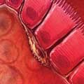
Macrophage
 Additional Markers
- Secreted
- TGF-beta 1.2
- TGF-beta 1/1.2
- TGF-beta 2
- TGF-beta 2/1.2
- TGF-beta 3
- TGF-beta 5
- Cell Surface
- CD11c
- CD68/SR-D1
- CD163
- Connexin 43/GJA1
- CX3CR1
- F4/80/EMR1
- Fc gamma RI/ CD64
- HLA-DR
- Integrin alpha M beta 2
- Integrin alpha M/CD11b Activators
- Langerin/CD207
- MHC class II
(I-A/I-E) - TLR10
- TREM-2
- Intracellular
- KDM6B
- KDM6B Inhibitors
- NF kappa B1
- NF kappa B1 Inhibitors
- TLR3
- TLR3 Inhibitors
- TLR3 Agonists
- TLR9
Phenotyping
Mouse
- CD68/SR-D1+
- CD163+
- F4/80/EMR1+
- IL-10+
- CD3-
- CD11c-
- CD19-
- IL-12low
Phenotyping
Phenotyping
Macrophage

Additional Markers
- Secreted
- EN-RAGE/ S100A12
- IL-12
- IL-23
- Cell Surface
- CD11c
- CX3CR1
- F4/80/EMR1
- Ly6C
- TLR10
- Intracellular
- PPAR gamma/ NR1C3
- PPAR gamma Agonists
- PPAR gamma Antagonists
- Additional PPAR gamma Products
- SOCS-3
- TLR3
- TLR3 Inhibitors
- TLR3 Agonists
Neutrophil
 Additional Markers
- Secreted
- alpha-Defensin 1
- APRIL/TNFSF13
- Azurocidin/CAP37/HBP
- BAFF/BLyS/TNFSF13B
- CAMP/LL37/FALL39
- CCL19/MIP-3 beta
- CCL2/JE/MCP-1
- CCL20/MIP-3 alpha
- CCL3/MIP-1 alpha
- CRISP-3
- iNOS
- iNOS Inhibitors
- Leukotrienes
- LIGHT/TNFSF14
- MMP-9
- MMP-9/Lipocalin-2 Complex
- MMP-9/TIMP-1 Complex
- MMP-9/TIMP-2 Complex
- MMP-9/TIMP-4 Complex
- Myeloperoxidase/MPO
- Neutrophil Elastase/ELA2
- Neutrophil Elastase/ELA2 Inhibitors
- Proteinase 3/ Myeloblastin/PRTN3
- ROS
- S100A Peptides
- VEGF
- Cell Surface
- AMICA/JAML
- CEACAM-8/CD66b
- Complement Component C3a R
- CXCR1/IL-8 RA
- CXCR2/IL-8 RB
- CXCR2/IL-8 RB Antagonists
- Dectin-1/CLEC7A
- Fc gamma RI/ CD64
- Fc gamma RIIA/ CD32a
- Fc gamma RIIIB/ CD16b
- FCAR/CD89
- FPR1
- FPR1 Agonists
- IL-23 R
- Integrin alpha M beta 2
- Integrin alpha M beta 2 Activators
- Integrin alpha X beta 2
- PSGL-1/CD162
- SIRP alpha/ CD172a
- TLR1
- TLR1 Inhibitors
- TLR2
- TLR2 Inhibitors
- TLR4
- TLR4 Inhibitors
- TLR4/MD-2 Complex
- TLR5
- TLR6
- TLR8
- TREM-1
- Intracellular
- NOD2
- TLR3
- TLR3 Agonists
- TLR3 Inhibitors
- TLR9
Phenotyping
- CEACAM-8/CD66b+
- Fc gamma RIII/CD16+
- Integrin alpha M/CD11b+
- Integrin beta 2/CD18+
- L-Selectin/CD62L+
- Myeloperoxidase+
- CD3-
- CD19-
Monocyte
 Additional Markers
- Secreted
- IL-1 alpha/IL-1F1
- IL-1 beta/IL-1F2
- IL-6
- Nitric oxide
- ROS
- TNF-alpha
- Cell Surface
- CCR2
- CCR2 Antagonists
- CD14
- FCAR/CD89
- Integrin alpha M/ CD11b
- Siglec-3/CD33
- Intracellular
- TLR9
Phenotyping
Eosinophil
 Additional Markers
- Secreted
- CCL5/RANTES
- CCL11/Eotaxin
- ECP/RNASE3
- EDN/RNASE2
- EPX
- GM-CSF
- IFN-gamma
- IL-2
- IL-4
- IL-5
- IL-5 R Antagonist
- IL-6
- IL-13
- MBP
- TGF-alpha
- TNF-alpha
- Cell Surface
- CCR3
- CCR3 Antagonists
- CD69
- F4/80/EMR1
- FCAR/CD89
- Integrin alpha 4/ CD49d
- Integrin alpha 4/ CD49d Inhibitors
- Integrin alpha M/ CD11b
- Integrin alpha M/ CD11b Activators
- Integrin beta 2/ CD18
- Siglec-8
- SIRP alpha/ CD172a
Phenotyping
Human
CX3CR1+ Dendritic Cell
 Additional Markers
- Secreted
- IL-6
- IL-12
- IL-23
- Osteopontin/OPN
- Cell Surface
- CD11c
- Connexin 43/GJA1
- CX3CR1
- DC-SIGN/CD209
- F4/80/EMR1
- Intracellular
- BATF3
Phenotyping
Human
- CD11c+
- CX3CR1+
- CD3-
- CD14-
- CD19-
- MS4A1/CD20-
- NCAM-1/CD56-
Mouse
- CX3CR1+
- F4/80/EMR1+
- CD3-
- CD11c-
- CD14-
- CD19-
- MS4A1/CD20-
- NCAM-1/CD56-
T Follicular Helper Cell (Tfh)
 Additional Markers
- Secreted
- TGF-beta 1.2
- TGF-beta 1/1.2
- TGF-beta 2
- TGF-beta 2/1.2
- TGF-beta 3
- TGF-beta 5
- Cell Surface
- BTLA
- CCR7
- CD3
- CD3 epsilon
- CD4
- CD28
- CD40 Ligand/ TNFSF5
- CD69
- CD84/SLAMF5
- CTLA-4
- CXCR5
- ICOS
- IL-10 R beta
- NTB-A/SLAMF6
- OX40/TNFRSF4
- PD-1
- SLAM/CD150
- Intracellular
- BATF
- Bcl-2
- Bcl-6
- c-Maf
- c-Rel
- IRF4
- NF kappa B1
- SH2D1A
- STAT1
- STAT3
- STAT4
Phenotyping
- Bcl-6+
- CD3+
- CD4+
- CXCR5+
- ICOS+
- PD-1+
- CD11c-
- CD19-
- CD25/IL-2 R alpha-
- GITR/TNFRSF18-
T Follicular Regulatory Cell (Tfr)
 Additional Markers
- Secreted
- TGF-beta 1.2
- TGF-beta 1/1.2
- TGF-beta 2
- TGF-beta 2/1.2
- TGF-beta 3
- TGF-beta 5
- Cell Surface
- BTLA
- CD3
- CD3 epsilon
- CD4
- CD25/IL-2 R alpha
- CD28
- CD44
- CTLA-4
- CXCR5
- GITR/TNFRSF18
- ICOS
- Integrin alpha E/ CD103
- KLRG1
- PD-1
- Intracellular
- Bcl-6
- BLIMP1/PRDM1
- FoxP3
- Helios
- SH2D1A
T Regulatory Cell (Treg)
 Additional Markers
- Secreted
- TGF-beta 1.2
- TGF-beta 1/1.2
- TGF-beta 2
- TGF-beta 2/1.2
- TGF-beta 3
- TGF-beta 5
- Cell Surface
- CCR9
- CD3
- CD3 epsilon
- CD4
- CD25/IL-2 R alpha
- CTLA-4
- GITR/TNFRSF18
- ICOS
- Integrin alpha 4 beta 7/ LPAM-1
- Integrin alpha 4/ CD49d
- Integrin alpha 4/ CD49d Inhibitors
- Integrin alpha E/ CD103
- Integrin beta 7
- Neuropilin-1
- TLR2
- TLR2 Inhibitors
- TLR4
- TLR4 Inhibitors
- TLR4/MD-2 Complex
- TLR5
- TLR7
- TLR7 Agonists
- TLR8
- TLR10
- TSLP R
- Intracellular
- FoxP3
- Helios
- ID2
- ID3
- STAT5a/b
- TLR9
Phenotyping
- CD3+
- CD4+
- CD25/IL-2 R alpha+
- FoxP3+
- GITR/TNFRSF18+
- CD11c-
- CD19-
Th17 Cell
 Additional Markers
- Secreted
- IL-6
- IL-9
- IL-10
- IL-17/IL-17A
- IL-17A/ F
Heterodimer - IL-17F
- IL-21
- IL-22
- IL-26/AK155
- LIGHT/TNFSF14
- TNF-alpha
- Cell Surface
- CCR4
- CCR6
- CD3
- CD3 epsilon
- CD4
- CD161
- CD161/NK1.1
- Common gamma Chain/IL-2 R gamma
- CRTAM
- gp130
- IL-1 RAcP/IL-1 R3
- IL-1 RI
- IL-6 R alpha
- IL-12 R beta 1
- IL-21 R
- IL-23 R
- TGF-beta RI/
ALK-5 - TGF-beta RII
- Intracellular
- AHR
- BATF
- IRF4
- ROR alpha/NR1F1
- ROR gamma t/ RORC2/NR1F3
- STAT1
- STAT3
- STAT4
- STAT5a/b
Phenotyping
- CCR4+
- CCR6+
- CD3+
- CD4+
- IL-6 R alpha+
- IL-12 R beta 1+
- IL-17+
- IL-23 R+
- ROR gamma t+
- CD11c-
- CD19-
Th9 Cell
 Additional Markers
- Secreted
- IL-9
- IL-10
- IL-21
- Cell Surface
- CCR3
- CCR3 Antagonists
- CCR6
- CD3
- CD3 epsilon
- CD4
- CXCR3
- IL-17 RB
- OX40/TNFRSF4
- Intracellular
- BATF
- GATA-3
- IRF4
- PU.1/Spi-1
- Smad3
- STAT6
Th22 Cell
 Additional Markers
- Secreted
- CCL15/
MIP-1 delta - CCL17/TARC
- CCL23/
Ck beta 8-1 - CCL23/MPIF-1
- IL-10
- IL-13
- IL-22
- TNF-alpha
- Cell Surface
- CCR4
- CCR6
- CCR10
- CD3
- CD3 epsilon
- CD4
- TSLP R
- Intracellular
- AHR
- BNC-2
- FoxO4
- ROR gamma/ RORC/NR1F3
- T-bet/TBX21
Tissue Resident Memory Cells (Trm)
 Additional Markers
- Secreted
- Granzyme B
- Granzyme B substrate
- Cell Surface
- CD69
- IL-2 R beta
- Integrin alpha E/ CD103
- L-Selectin/CD62L
- Ly-6C
- Intracellular
- EOMES
Phenotyping
- CD8+
- CD69+
- Granzyme B+
- IL-12 R beta 1+
- Integrin alpha E/CD103+
- Ly-6Clow
- L-Selectin/CD62Llow
- CD11c-
- CD19-
Regulatory NK Cell
 Additional Markers
- Secreted
- IFN-gamma
- IL-17/IL-17A
- IL-17A/ F
Heterodimer - IL-22
- Cell Surface
- CD117/c-kit
- IL-17 RB
- NCAM-1/CD56
- TLR2
- TLR2 Inhibitors
- Intracellular
- TLR3
- TLR3 Inhibitors
- TLR3 Agonists
- TLR9
Phenotyping
Lti Cell
 Additional Markers
- Secreted
- IL-17/IL-17A
- IL-17A/ F
Heterodimer - IL-22
- Lymphotoxin-alpha/TNF-beta
- Lymphotoxin beta/TNFSF3
- Cell Surface
- CCR6
- CD30 Ligand/ TNFSF8
- CD117/c-kit
- Common gamma Chain/IL-2 R gamma
- IL-1 RI
- IL-2 R beta
- IL-7 R alpha/ CD127
- IL-23 R
- OX40 Ligand/ TNFSF4
- TLR2
- TLR2 Inhibitors
- TLR4
- TLR4 Inhibitors
- TLR4/MD-2 Complex
- TRANCE/TNFSF11/
RANK L - TSLP R
- Intracellular
- ROR gamma t/ RORC2/NR1F3
- TCF7/TCF1
Phenotyping
ILC2
 Additional Markers
- Secreted
- GM-CSF
- IFN-gamma
- IL-2
- IL-17/IL-17A
- IL-17A/ F
Heterodimer - IL-22
- Lymphotoxin beta/TNFSF3
- Lymphotoxin-alpha/TNF-beta
- TNF-alpha
- Cell Surface
- 2B4/CD244/SLAMF4
- CCR6
- CCR7
- CD117/c-Kit
- CD161
- CD161/NK1.1
- Common gamma Chain/IL-2 R gamma
- CXCR6
- DNAM-1/CD226
- IL-2 R beta
- IL-7 R alpha/ CD127
- IL-23 R
- NCAM-1/CD56
- NKp30/NCR3
- NKp44/NCR2
- NKp46/NCR1
- TRANCE/TNFSF11/
RANK L - TSLP R
- Intracellular
- AHR
- GATA-3
- ROR gamma t/ RORC2/NR1F3
Phenotyping
Phenotyping
Human
- CD117/c-kit+
- IL-7 R alpha/ CD127+
- IL-23 R+
- ROR gamma t+
- CD4+/-
- CD94+/-
- NKG2D+/-
- NKp46+/-
- CD11c-
- CD19-
Mouse
- CD117/c-kit+
- IL-7 R alpha/ CD127+
- IL-23 R+
- ROR gamma t+
- T-bet+
- CD4+/-
- CD94+/-
- NKG2D+/-
- NKp46+/-
- CD161low
- NCAM-1/CD56low
- NKp30/NCR3low
- NKp44/NCR2low
- CD11c-
- CD19-
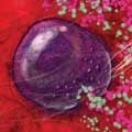
Cytotoxic NK Cell
 Additional Markers
- Secreted
- Granzyme A
- Granzyme B
- Granzyme H
- IFN-gamma
- Perforin
- TNF-alpha
- Cell Surface
- CCR2
- CCR2 Antagonists
- CCR5
- CCR5 Antagonists
- CCR7
- CD94
- CD117/c-Kit
- CX3CR1
- CXCR1/IL-8 RA
- DNAM-1/CD226
- Fc gamma RIIIA/ CD16a
- Fc gamma RIIIB/ CD16b
- NCAM-1/CD56
- NKG2D/CD314
- NKp30/NCR3
- NKp44/NCR2
- NKp46/NCR1
- TLR1
- TLR1 Agonists
- TLR1 Inhibitors
- TLR2
- TLR2 Agonists
- TLR2 Inhibitors
- TLR4
- TLR4 Inhibitors
- TLR4/
MD-2 Complex - TLR5
- TLR6
- TLR7
- TLR7 Agonists
- TLR8
- Intracellular
- BLIMP1/PRDM1
- EOMES
- Helios
- T-bet/TBX21
- TLR3
- TLR3 Agonists
- TLR3 Inhibitors
- ZBTB32
Phenotyping
Phenotyping
Human
- CD117/c-kit+
- IL-7 R alpha/ CD127+
- IL-23 R+
- ROR gamma t+
- CD4+/-
- CD94+/-
- NKG2D+/-
- NKp46+/-
- CD11c-
- CD19-
Mouse
- CD68/SR-D1+
- CD117/c-kit+
- CD163+
- F4/80/EMR1+
- IL-7 R alpha/ CD127+
- IL-10+
- IL-23 R+
- ROR gamma t+
- T-bet+
- CD4+/-
- CD94+/-
- NKG2D+/-
- NKp46+/-
- CD161low
- NCAM-1/CD56low
- NKp30low
- NKp44low
- CD11c-
- CD19-
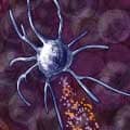
Follicular Dendritic Cell (FDC)
Additional Markers
- Secreted
- TGF-beta 1.2
- TGF-beta 1/1.2
- TGF-beta 2
- TGF-beta 2/1.2
- TGF-beta 3
- TGF-beta 5
- Wnt-5a
- Cell Surface
- CD21
- CD35
- CD36/SR-B3
- DLL1
- Fc gamma RII/ CD32
- Fc gamma RII/ RIII (CD32/CD16)
- Fc gamma RIIA/ CD32a
- Fc gamma RIIB/
C (CD32b/c) - Fc gamma RIIB/ CD32b
- Fc gamma RIIC/ CD32c
- Fc gamma RIIIA/ CD16a
- Fc gamma RIIIB/ CD16b
- HLA-DR
- ICAM-1/CD54
- ICAM-1/
CD54 Inhibitors - Jagged 1
- Jagged 1/
Jagged 2 - Lymphotoxin beta R/TNFRSF3
- MAdCAM-1
- MD-2
- MHC class II
(I-A/I-E) - Podoplanin
- Sonic Hedgehog/ Shh
- TLR2
- TLR2 Inhibitors
- TLR4
- TLR4 Inhibitors
- TLR4/
MD-2 Complex - TLR5
- VCAM-1/CD106
- Intracellular
- Aldehyde Dehydrogenase 1-A1/ALDH1A1
- ALDH1A2
- ALDH1A3
- IKK beta
- NF kappa B1
- TLR9
Phenotyping
- CD19+
- MS4A1/CD20+
- CD3-
- CD4-
- CD11c-
Phenotyping
- CD27/TNFRSF7+
- surface IgA+
- Syndecan-1/CD138+/-
- CD19low
- CD3-
- CD4-
- CD11c-
- MS4A1/CD20-
Phenotyping
- CD38high
- Intracellular IgA+
- Syndecan-1/CD138+
- CD19low
- CD3-
- CD4-
- CD11c-
- MS4A1/CD20-
Phenotyping
- CD38high
- Syndecan-1/CD138+
- CD19low
- CD3-
- CD4-
- CD11c-
- MS4A1/CD20-
Phenotyping
- CD2+
- CD5+
- CD8 alpha/beta+
- CD11c+
- CD28+
- CD44+
- CD90/Thy1+
- CTLA-4+
- Integrin alpha L/CD11a+
- TCR alpha/beta+
- CD4+/-
- CD19-
Phenotyping
Phenotyping
Overview of Intestinal Lamina Propria Immune Cells
Immune system function in the healthy intestine is tolerogenic. Dendritic cells and macrophages acquire commensal antigenic materials transported across the epithelium or directly from the intestinal lumen. These cells as well as lymphoid cells secrete immunoregulatory cytokines and other mediators that block the development of inflammatory responses and reinforce epithelial barrier integrity. Follicular dendritic cells and T cells promote the development of IgA-producing B cells in local Peyer’s patches. During epithelial breakdown or pathogen infection, homeostatic immune cell activity is suppressed. Inflammatory cells infiltrate the lamina propria to kill and clear invading microbes. The immune response is dominated by the release of proinflammatory cytokines, chemokines, proteases, and reactive oxygen species.

