Human Parkin Antibody Summary
Met1-Val437
Accession # NP_054642
Customers also Viewed
Applications
Please Note: Optimal dilutions should be determined by each laboratory for each application. General Protocols are available in the Technical Information section on our website.
Scientific Data
 View Larger
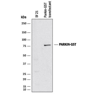
Detection of Human Parkin by Western Blot. Western blot shows lysates of Sf 21 S. frugiperda insect ovarian cell line mock transfected or transfected GST tagged Parkin. PVDF membrane was probed with 2 µg/mL of Goat Anti-Human Parkin Antigen Affinity-purified Polyclonal Antibody (Catalog # AF1438) followed by HRP-conjugated Anti-Goat IgG Secondary Antibody (Catalog # HAF017). A specific band was detected for GST tagged Parkin at approximately 75 kDa (as indicated). This experiment was conducted under reducing conditions and using Immunoblot Buffer Group 1.
 View Larger
Parkin in Human Brain. Parkin was detected in immersion fixed paraffin-embedded sections of human brain (cerebellum) using 15 µg/mL Goat Anti-Human Parkin Antigen Affinity-purified Polyclonal Antibody (Catalog # AF1438) overnight at 4 °C. Tissue was stained with the Anti-Goat HRP-DAB Cell & Tissue Staining Kit (brown; Catalog # CTS008) and counterstained with hematoxylin (blue). View our protocol for Chromogenic IHC Staining of Paraffin-embedded Tissue Sections.
Preparation and Storage
- 12 months from date of receipt, -20 to -70 °C as supplied.
- 1 month, 2 to 8 °C under sterile conditions after reconstitution.
- 6 months, -20 to -70 °C under sterile conditions after reconstitution.
Background: Parkin
Parkin is a 465 amino acid protein with an N-terminal ubiquitin-like domain, a linker region, a C-terminal TRIAD domain consisting of two RING finger motifs flanking a cysteine-rich IBR (in between RING fingers) motif. Mutations in Parkin is a major cause of autosomal recessive juvenile parkinsonism. Parkin-2 lacks exon 5 which encodes amino acid residues 179-206 of Parkin.
Product Datasheets
Citations for Human Parkin Antibody
R&D Systems personnel manually curate a database that contains references using R&D Systems products. The data collected includes not only links to publications in PubMed, but also provides information about sample types, species, and experimental conditions.
2
Citations: Showing 1 - 2
Filter your results:
Filter by:
-
Distinct phosphorylation signals drive acceptor versus free ubiquitin chain targeting by parkin
Authors: Karen M. Dunkerley, Anne C. Rintala-Dempsey, Giulia Salzano, Roya Tadayon, Dania Hadi, Kathryn R. Barber et al.
Biochemical Journal
-
Interleukin-1beta drives NEDD8 nuclear-to-cytoplasmic translocation, fostering parkin activation via NEDD8 binding to the P-ubiquitin activating site
Authors: M Balasubram, PA Parcon, C Bose, L Liu, RA Jones, MR Farlow, RE Mrak, SW Barger, WST Griffin
J Neuroinflammation, 2019-12-27;16(1):275.
Species: Human
Sample Types: Whole Tissue
Applications: IHC-P
FAQs
No product specific FAQs exist for this product, however you may
View all Antibody FAQsReviews for Human Parkin Antibody
There are currently no reviews for this product. Be the first to review Human Parkin Antibody and earn rewards!
Have you used Human Parkin Antibody?
Submit a review and receive an Amazon gift card.
$25/€18/£15/$25CAN/¥75 Yuan/¥2500 Yen for a review with an image
$10/€7/£6/$10 CAD/¥70 Yuan/¥1110 Yen for a review without an image


