Mouse ESAM Antibody Summary
Gln30-Ser248 (predicted)
Accession # Q925F2
*Small pack size (-SP) is supplied either lyophilized or as a 0.2 µm filtered solution in PBS.
Customers also Viewed
Applications
Please Note: Optimal dilutions should be determined by each laboratory for each application. General Protocols are available in the Technical Information section on our website.
Scientific Data
 View Larger
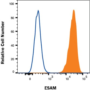
Detection of ESAM in bEnd.3 cells by Flow Cytometry bEnd.3 cells were stained with Goat Anti-Mouse ESAM Antigen Affinity-purified Polyclonal Antibody (Catalog # AF2827, filled histogram) or isotype control antibody (Catalog # AB-108-C, open histogram) followed by Allophycocyanin-conjugated Anti-Goat IgG Secondary Antibody (Catalog # F0108). View our protocol for Staining Membrane-associated Proteins.
 View Larger
Detection of ESAM in Mouse Kidney. ESAM was detected in immersion fixed paraffin-embedded sections of mouse kidney using Goat Anti-Mouse ESAM Antigen Affinity-purified Polyclonal Antibody (Catalog # af2827) at 5 µg/ml for 1 hour at room temperature followed by incubation with the Anti-Goat IgG VisUCyte™ HRP Polymer Antibody (Catalog # VC004). Before incubation with the primary antibody, tissue was subjected to heat-induced epitope retrieval using VisUCyte Antigen Retrieval Reagent-Basic (Catalog # VCTS021). Tissue was stained using DAB (brown) and counterstained with hematoxylin (blue). Specific staining was localized to the cell surface of vascular endothelial cells. View our protocol for IHC Staining with VisUCyte HRP Polymer Detection Reagents.
 View Larger
Detection of ESAM in Mouse Liver. ESAM was detected in immersion fixed paraffin-embedded sections of mouse liver using Goat Anti-Mouse ESAM Antigen Affinity-purified Polyclonal Antibody (Catalog # af2827) at 5 µg/ml for 1 hour at room temperature followed by incubation with the Anti-Goat IgG VisUCyte™ HRP Polymer Antibody (Catalog # VC004). Before incubation with the primary antibody, tissue was subjected to heat-induced epitope retrieval using VisUCyte Antigen Retrieval Reagent-Basic (Catalog # VCTS021). Tissue was stained using DAB (brown) and counterstained with hematoxylin (blue). Specific staining was localized to the cell surface of vascular endothelial cells. View our protocol for IHC Staining with VisUCyte HRP Polymer Detection Reagents.
Preparation and Storage
- 12 months from date of receipt, -20 to -70 °C as supplied.
- 1 month, 2 to 8 °C under sterile conditions after reconstitution.
- 6 months, -20 to -70 °C under sterile conditions after reconstitution.
Background: ESAM
ESAM is a 55 kDa type I transmembrane glycoprotein belonging to the CTX (cortical thymocyte marker in Xenopus) family of cell adhesion molecules within the immunoglobulin superfamily. Other family members are CXADR, CLMP, JAM-A-C, CD2, A33, and BT-IgSF. The extracellular region of ESAM contains one V-type and one C2-type Ig domain and is involved in homophilic adhesion. Mouse ESAM extracellular domain shares 69% amino acid sequence identity with the corresponding region of human ESAM. ESAM is expressed on endothelial cells, activated platelets and megakaryocytes and can be found associated with cell-to-cell junctions.
Product Datasheets
Citations for Mouse ESAM Antibody
R&D Systems personnel manually curate a database that contains references using R&D Systems products. The data collected includes not only links to publications in PubMed, but also provides information about sample types, species, and experimental conditions.
3
Citations: Showing 1 - 3
Filter your results:
Filter by:
-
Claudin5 protects the peripheral endothelial barrier in an organ and vessel-type-specific manner
Authors: Mark Richards, Emmanuel Nwadozi, Sagnik Pal, Pernilla Martinsson, Mika Kaakinen, Marleen Gloger et al.
eLife
-
A Prox1 enhancer represses haematopoiesis in the lymphatic vasculature
Authors: J Kazenwadel, P Venugopal, A Oszmiana, J Toubia, L Arriola-Ma, V Panara, SG Piltz, C Brown, W Ma, AW Schreiber, K Koltowska, S Taoudi, PQ Thomas, HS Scott, NL Harvey
Nature, 2023-01-25;614(7947):343-348.
Species: Mouse
Sample Types: Whole Tissue
Applications: IHC -
A RORgammat+ cell instructs gut microbiota-specific Treg cell differentiation
Authors: R Kedmi, TA Najar, KR Mesa, A Grayson, L Kroehling, Y Hao, S Hao, M Pokrovskii, M Xu, J Talbot, J Wang, J Germino, CA Lareau, AT Satpathy, MS Anderson, TM Laufer, I Aifantis, JM Bartleson, PM Allen, H Paidassi, JM Gardner, M Stoeckius, DR Littman
Nature, 2022-09-07;0(0):.
Species: Transgenic Mouse
Sample Types: Whole Cells
Applications: CITE-seq
FAQs
No product specific FAQs exist for this product, however you may
View all Antibody FAQsReviews for Mouse ESAM Antibody
There are currently no reviews for this product. Be the first to review Mouse ESAM Antibody and earn rewards!
Have you used Mouse ESAM Antibody?
Submit a review and receive an Amazon gift card.
$25/€18/£15/$25CAN/¥75 Yuan/¥2500 Yen for a review with an image
$10/€7/£6/$10 CAD/¥70 Yuan/¥1110 Yen for a review without an image


