Human B-Raf Antibody Summary
Met1-His766
Accession # P15056
Applications
Please Note: Optimal dilutions should be determined by each laboratory for each application. General Protocols are available in the Technical Information section on our website.
Scientific Data
 View Larger
Detection of Human B-Raf by Western Blot. Western blot shows lysates of T47D human breast cancer cell line, MDA-MB-453 human breast cancer cell line, Daudi human Burkitt's lymphoma cell line, MOLT-4 human acute lymphoblastic leukemia cell line and U266 human myeloma cell line. PVDF membrane was probed with 1 µg/mL of Goat Anti-Human B-Raf Antigen Affinity-purified Polyclonal Antibody (Catalog # AF3424) followed by HRP-conjugated Anti-Goat IgG Secondary Antibody (Catalog # HAF109). A specific band was detected for B-Raf at approximately 95 kDa (as indicated). This experiment was conducted under reducing conditions and using Immunoblot Buffer Group 1.
 View Larger
Detection of Human B‑Raf by Simple WesternTM. Simple Western lane view shows lysates of T47D human breast cancer cell line, loaded at 0.2 mg/mL. A specific band was detected for B-Raf at approximately 95 kDa (as indicated) using 10 µg/mL of Goat Anti-Human B-Raf Antigen Affinity-purified Polyclonal Antibody (Catalog # AF3424) followed by 1:50 dilution of HRP-conjugated Anti-Goat IgG Secondary Antibody (Catalog # HAF109). This experiment was conducted under reducing conditions and using the 12-230 kDa separation system.
 View Larger
Detection of Human B‑Raf by Simple WesternTM. Simple Western lane view shows lysates of MOLT‑4 human acute lymphoblastic leukemia cell line, loaded at 0.2 mg/mL. A specific band was detected for B‑Raf at approximately 95 kDa (as indicated) using 10 µg/mL of Goat Anti-Human B‑Raf Antigen Affinity-purified Polyclonal Antibody (Catalog # AF3424) followed by 1:50 dilution of HRP-conjugated Anti-Goat IgG Secondary Antibody (Catalog # HAF109). This experiment was conducted under reducing conditions and using the 12-230 kDa separation system.
 View Larger
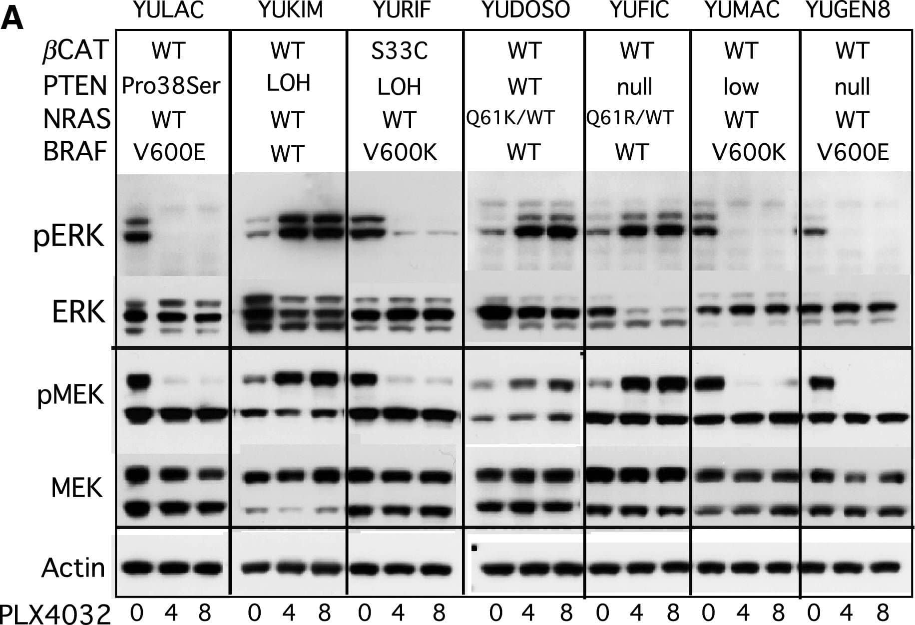
Detection of Human B-Raf by Western Blot Changes in ERK1/2 and MEK in response to PLX4032. (A) Melanoma cell strains were treated with DMSO for 4 h (0), or with PLX4032 (1 μM) for 4 and 8 h. The panels show Western blots probed with antibodies to phosph-ERK1/2 Thr202/Tyr204 mAb (pERK), ERK1/2 (ERK), phospho-MEK1/2 (pMEK), MEK1/2 (MEK), and actin as a loading control. The mutation status of BRAF, NRAS, PTEN and beta -catenin ( beta CAT) are indicated at the top. (B) Western blot analyses of ERK1/2 inactivation/activation after short-term incubation with PLX4032 (1 μM). (C) pERK1/2 and ERK1/2 in supernatant (Sup) and particulate (Part) fractions. (D) Changes in pERK1/2 activation after treatments with increasing concentration of PLX4032, or DMSO for 1 h. Image collected and cropped by CiteAb from the following open publication (https://pubmed.ncbi.nlm.nih.gov/20149136), licensed under a CC-BY license. Not internally tested by R&D Systems.
Preparation and Storage
- 12 months from date of receipt, -20 to -70 °C as supplied.
- 1 month, 2 to 8 °C under sterile conditions after reconstitution.
- 6 months, -20 to -70 °C under sterile conditions after reconstitution.
Background: B-Raf
The Raf serine/threonine kinases are effectors of Ras that function as MAP3Ks in the ERK phosphorylation cascade. Mammals express three Raf proteins: Raf-1 (C‑Raf); A-Raf; and B-Raf, found at high levels in cerebrum and testes. Mice with a targeted disruption of the B-Raf gene die of vascular defects during mid-gestation. B-Raf mutations have been found in two-thirds of malignant melanomas, with the single substitution V599E in the kinase domain the most frequent occurrence. B-Raf activation in melanomas results in BIM phosphorylation and inhibition of apoptosis.
Product Datasheets
Citation for Human B-Raf Antibody
R&D Systems personnel manually curate a database that contains references using R&D Systems products. The data collected includes not only links to publications in PubMed, but also provides information about sample types, species, and experimental conditions.
1 Citation: Showing 1 - 1
-
PLX4032, a selective BRAF(V600E) kinase inhibitor, activates the ERK pathway and enhances cell migration and proliferation of BRAF melanoma cells.
Authors: Halaban R, Zhang W, Bacchiocchi A et al.
Pigment Cell Melanoma Res
FAQs
No product specific FAQs exist for this product, however you may
View all Antibody FAQsReviews for Human B-Raf Antibody
Average Rating: 4 (Based on 1 Review)
Have you used Human B-Raf Antibody?
Submit a review and receive an Amazon gift card.
$25/€18/£15/$25CAN/¥75 Yuan/¥2500 Yen for a review with an image
$10/€7/£6/$10 CAD/¥70 Yuan/¥1110 Yen for a review without an image
Filter by:


