Human CD34 Antibody Summary
Ser32-Thr290
Accession # P28906-1
Applications
Please Note: Optimal dilutions should be determined by each laboratory for each application. General Protocols are available in the Technical Information section on our website.
Scientific Data
 View Larger
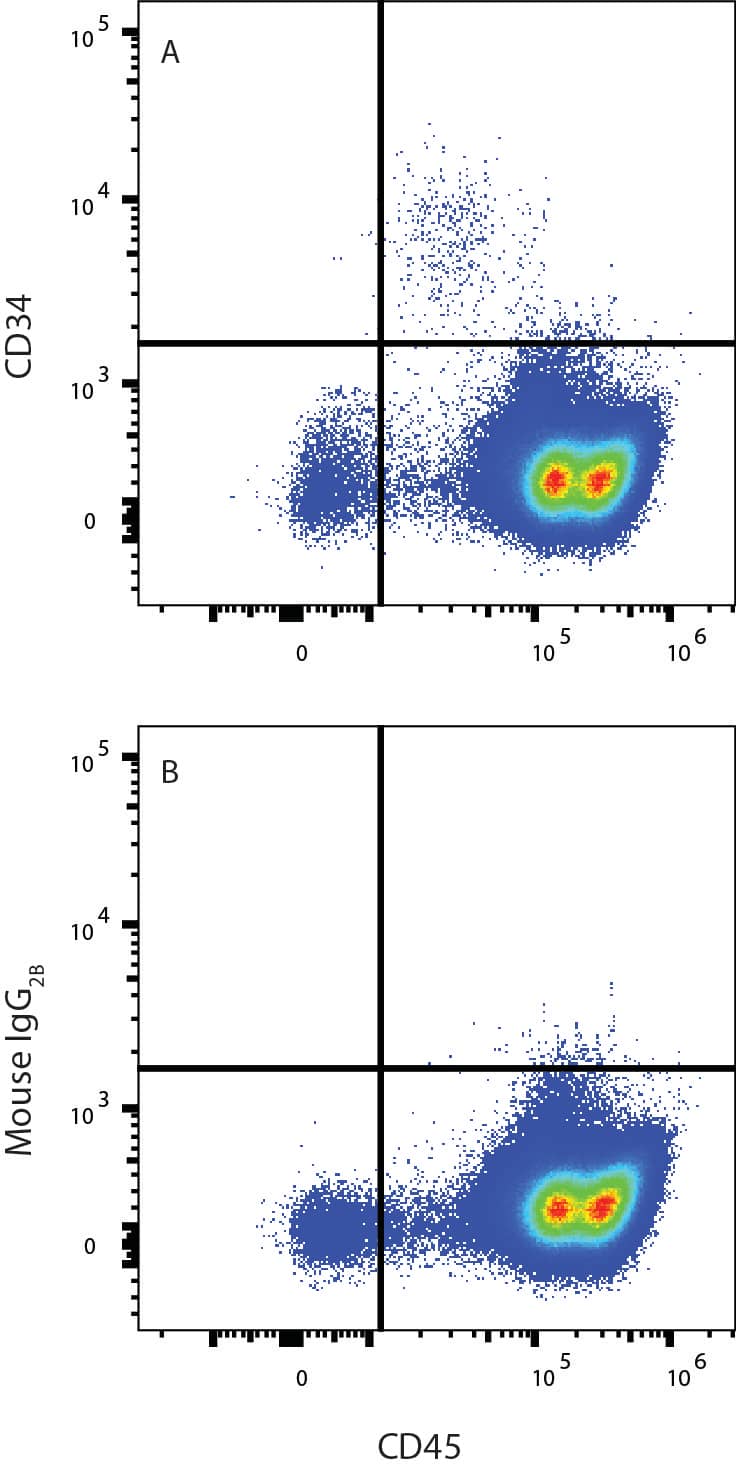
Detection of CD34 in Human PBMCs by Flow Cytometry. Human peripheral blood mononuclear cells (PBMCs) were stained with (A) Mouse Anti-Human CD34 Monoclonal Antibody (Catalog # MAB72274) or (B) Mouse IgG2B isotype control antibody (MAB0041) followed by APC-conjugated Anti-Mouse IgG secondary antibody and Mouse Anti-Human CD45 PE-conjugated Monoclonal Antibody (FAB1430P). View our protocol for Staining Membrane-associated Proteins.
 View Larger
Detection of CD34 in KG-1 (positive) and K562 (negative) cells. CD34 was detected in immersion fixed KG-1 (positive) and absent in K562 (negative) cells using Mouse Anti-Human CD34 Monoclonal Antibody (Catalog # MAB72274) at 8 µg/mL for 3 hours at room temperature. Cells were stained using the NorthernLights™ 557-conjugated Anti-Mouse IgG Secondary Antibody (red; Catalog # NL007) and counterstained with DAPI (blue). Specific staining was localized to cytoplasm. View our protocol for Fluorescent ICC Staining of Non-adherent Cells.
 View Larger
Detection of CD34 in KG-1 cells by Flow Cytometry. KG-1 cells were stained with Mouse Anti-Human CD34 Monoclonal Antibody (Catalog # MAB72274, filled histogram) or isotype control antibody (Catalog # MAB0041, open histogram), followed by Phycoerythrin-conjugated Anti-Mouse IgG Secondary Antibody (Catalog # F0102B). View our protocol for Staining Membrane-associated Proteins.
Reconstitution Calculator
Preparation and Storage
- 12 months from date of receipt, -20 to -70 °C as supplied.
- 1 month, 2 to 8 °C under sterile conditions after reconstitution.
- 6 months, -20 to -70 °C under sterile conditions after reconstitution.
Background: CD34
CD34 is a 115 kDa glycosylated type I transmembrane protein; it was discovered as a hematopoietic cell-surface antigen (1, 2, 3). Human CD33 cDNA encodes a 385 amino acid (aa) precursor that contains a 31 aa signal sequence, a 259 aa extracellular domain (ECD), a 21 aa transmembrane sequence, and a 74 aa cytoplasmic domain. Within the ECD, human CD34 shares 55% and 52% aa sequence identity with mouse and rat CD34, respectively. This single-pass sialomucin‑like transmembrane protein is heavily glycosylated and phosphorylated by Protein Kinase C (PKC) (4, 5). CD34 is found on multipotent precursors, bone marrow stromal cells, embryonic fibroblasts, vascular endothelia, as well as some populations of mesenchymal stem cells, and tumor cell lines, and it is a common marker for diverse progenitors (6). CD34 is involved in the adhesion of stem cells to the bone marrow extracellular matrix or to stromal cells.
- Civin C.I. et al. (1984) J. Immunol. 133:157.
- Katz F. et al. (1985) Leuk. Res. 9:191.
- Andrews R.G. et al. (1986) Blood 67:842.
- Young P.E. et al. (1995) Blood 85:96.
- Krause D.S. et al. (1996) Blood 87:1.
- Sidney L.E. et al. (2014) Stem Cells 32:1380.
Product Datasheets
FAQs
No product specific FAQs exist for this product, however you may
View all Antibody FAQsReviews for Human CD34 Antibody
There are currently no reviews for this product. Be the first to review Human CD34 Antibody and earn rewards!
Have you used Human CD34 Antibody?
Submit a review and receive an Amazon gift card.
$25/€18/£15/$25CAN/¥75 Yuan/¥2500 Yen for a review with an image
$10/€7/£6/$10 CAD/¥70 Yuan/¥1110 Yen for a review without an image


