Human CD34 Antibody Summary
Ser32-Thr290
Accession # P28906
Applications
Please Note: Optimal dilutions should be determined by each laboratory for each application. General Protocols are available in the Technical Information section on our website.
Scientific Data
 View Larger
Detection of Human CD34 by Western Blot. Western blot shows lysates of human testies tissue and human thymus tissue. PVDF membrane was probed with 0.5 µg/mL of Sheep Anti-Human CD34 Antigen Affinity-purified Polyclonal Antibody (Catalog # AF7227) followed by HRP-conjugated Anti-Sheep IgG Secondary Antibody (Catalog # HAF016). A specific band was detected for CD34 at approximately 110 kDa (as indicated). This experiment was conducted under reducing conditions and using Immunoblot Buffer Group 1.
 View Larger
CD34 in Human Liver. CD34 was detected in immersion fixed paraffin-embedded sections of human liver using Sheep Anti-Human CD34 Antigen Affinity-purified Polyclonal Antibody (Catalog # AF7227) at 1.7 µg/mL overnight at 4 °C. Tissue was stained using the Anti-Sheep HRP-DAB Cell & Tissue Staining Kit (brown; Catalog # CTS019) and counterstained with hematoxylin (blue). Specific staining was localized to endothelial cells in vasculature. View our protocol for Chromogenic IHC Staining of Paraffin-embedded Tissue Sections.
 View Larger
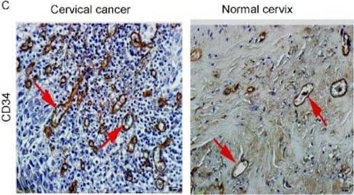
Detection of Human CD34 by Immunohistochemistry sAng-2 concentration positively relates with Ang-2 expression on the epithelia and MVD in cervical tissues.Representative immunohistochemical staining of Ang-1 (A), Ang-2 (B) and CD34 (C) in 25 cervical cancer tissue specimens and 10 normal controls. Black arrows denote positively stained epithelial cells, whereas red arrows denote positively staining endothelial cells, all appearing brown. Scale bar, 20 µm. (D) sAng-2 is significantly higher in the patients with positive Ang-2 expression on cervix epithelia than those with negative Ang-2 expression. The scatter diagrams show the correlations of sAng-1 (E), sAng-2 (F) and sAng-1/ sAng-2 ratio (G) to MVD in the 35 cervical tissue specimens. Image collected and cropped by CiteAb from the following publication (https://pubmed.ncbi.nlm.nih.gov/28584715), licensed under a CC-BY license. Not internally tested by R&D Systems.
Preparation and Storage
- 12 months from date of receipt, -20 to -70 °C as supplied.
- 1 month, 2 to 8 °C under sterile conditions after reconstitution.
- 6 months, -20 to -70 °C under sterile conditions after reconstitution.
Background: CD34
CD34 is a 105-115 kDa member of the CD34/podocalyxin family of molecules. It is a sialomucin type glycoprotein, and presents carbohydrate to selectins during cell migration. CD34 is found on mast cells, eosinophils, vascular endothelial cells, stem cells and renal mesangial cells. Mature human CD34 is a 354 amino acid (aa) type I transmembrane protein (aa 32-385). It contains a 259 aa extracellular region (aa 35-287) with utilized N- and O-linked glycosylation sites, and a 74 aa cytoplasmic domain that may undergo Tyr phosphorylation. There is one splice variant that shows a four aa substitution for aa 325-385. Human CD34 can undergo membrane cleavage by bacterial proteases to generate 30-40 kDa soluble fragments. And notably, desialylated CD34 shows a 40 kDa increase in MW (to 150 kDa) when run in SDS-PAGE. Over aa 32-290, human CD34 shares 56% aa identity with mouse CD34.
Product Datasheets
Citations for Human CD34 Antibody
R&D Systems personnel manually curate a database that contains references using R&D Systems products. The data collected includes not only links to publications in PubMed, but also provides information about sample types, species, and experimental conditions.
8
Citations: Showing 1 - 8
Filter your results:
Filter by:
-
Single-Cell RNA Sequencing Unifies Developmental Programs of Esophageal and Gastric Intestinal Metaplasia
Authors: Karol Nowicki-Osuch, Lizhe Zhuang, Tik Shing Cheung, Emily L. Black, Neus Masqué-Soler, Ginny Devonshire et al.
Cancer Discovery
-
Three-dimensional imaging of upper tract urothelial carcinoma improves diagnostic yield and accuracy
Authors: Fukumoto, K;Kanatani, S;Jaremko, G;West, Z;Li, Y;Takamatsu, K;Al Rayyes, I;Mikami, S;Niwa, N;Axelsson, TA;Tanaka, N;Oya, M;Miyakawa, A;Brehmer, M;Uhlén, P;
JCI insight
Species: Mouse
Sample Types: Whole Tissue
Applications: Immunohistochemistry -
NOTCH3 drives meningioma tumorigenesis and resistance to radiotherapy
Authors: Choudhury, A;Cady, M;Lucas, C;Najem, H;Phillips, J;Palikuqi, B;Zakimi, N;Joseph, T;Birrueta, J;Chen, W;Bush, N;Hervey-Jumper, S;Klein, O;Toedebusch, C;Horbinski, C;Magill, S;Bhaduri, A;Perry, A;Dickinson, P;Heimberger, A;Ashworth, A;Crouch, E;Raleigh, D;
bioRxiv
Species: Human
Sample Types: Whole Tissue
Applications: IHC -
An open source toolkit for repurposing Illumina sequencing systems as versatile fluidics and imaging platforms
Authors: K Pandit, J Petrescu, M Cuevas, W Stephenson, P Smibert, H Phatnani, S Maniatis
Scientific Reports, 2022-03-24;12(1):5081.
Species: Human
Sample Types: Whole Tissue
Applications: IHC -
Reciprocal Interaction between Vascular Filopodia and Neural Stem Cells Shapes Neurogenesis in the Ventral Telencephalon
Authors: B Di Marco, EE Crouch, B Shah, C Duman, MF Paredes, C Ruiz de Al, EJ Huang, J Alfonso
Cell Rep, 2020-10-13;33(2):108256.
Species: Human
Sample Types: Whole Tissue
Applications: IHC -
Multicolor quantitative confocal imaging cytometry
Authors: DL Coutu, KD Kokkaliari, L Kunz, T Schroeder
Nat. Methods, 2017-11-13;15(1):39-46.
Species: Mouse
Sample Types: Whole Tissue
Applications: IHC -
Effects of negative-pressure wound therapy combinedwith microplasma on treating wounds of ulcer and the expression of heat shock protein 90
Authors: Z Li, Q Wang, W Mi, M Han, F Gao, G Niu, Y Ma
Exp Ther Med, 2017-03-27;13(5):2211-2216.
Species: Human
Sample Types: Whole Tissue
Applications: IHC-P -
A human cell atlas of fetal gene expression
Authors: Cao J, O'Day DR, Pliner HA et al.
Science
FAQs
No product specific FAQs exist for this product, however you may
View all Antibody FAQsReviews for Human CD34 Antibody
Average Rating: 5 (Based on 1 Review)
Have you used Human CD34 Antibody?
Submit a review and receive an Amazon gift card.
$25/€18/£15/$25CAN/¥75 Yuan/¥2500 Yen for a review with an image
$10/€7/£6/$10 CAD/¥70 Yuan/¥1110 Yen for a review without an image
Filter by:


