Human CKAP4/p63 Antibody Summary
His128-Val602
Accession # Q07065
Applications
Please Note: Optimal dilutions should be determined by each laboratory for each application. General Protocols are available in the Technical Information section on our website.
Scientific Data
 View Larger
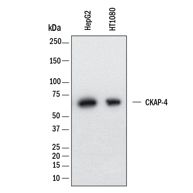
Detection of Human CKAP4/p63 by Western Blot. Western Blot shows lysates of HepG2 human hepatocellular carcinoma cell line and HT1080 human fibrosarcoma cell line. PVDF membrane was probed with 1 µg/ml of Mouse Anti-Human CKAP4/p63 Monoclonal Antibody (Catalog # MAB11600) followed by HRP-conjugated Anti-Mouse IgG Secondary Antibody (Catalog # HAF018). A specific band was detected for CKAP4/p63 at approximately 65 kDa (as indicated). This experiment was conducted under reducing conditions and using Western Blot Buffer Group 1.
 View Larger
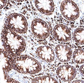
Detection of CKAP4/p63 in Human Colon. CKAP4/p63 was detected in immersion fixed paraffin-embedded sections of human colon using Mouse Anti-Human CKAP4/p63 Monoclonal Antibody (Catalog # MAB11600) at 5 µg/ml for 1 hour at room temperature followed by incubation with the Anti-Mouse IgG VisUCyte™ HRP Polymer Antibody (Catalog # VC001). Before incubation with the primary antibody, tissue was subjected to heat-induced epitope retrieval using VisUCyte Antigen Retrieval Reagent-Basic (Catalog # VCTS021). Tissue was stained using DAB (brown) and counterstained with hematoxylin (blue). Specific staining was localized to the cell surface and cytoplasm. View our protocol for IHC Staining with VisUCyte HRP Polymer Detection Reagents.
 View Larger
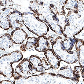
Detection of CKAP4/p63 in Human Placenta. CKAP4/p63 was detected in immersion fixed paraffin-embedded sections of human placenta using Mouse Anti-Human CKAP4/p63 Monoclonal Antibody (Catalog # MAB11600) at 5 µg/ml for 1 hour at room temperature followed by incubation with the Anti-Mouse IgG VisUCyte™ HRP Polymer Antibody (Catalog # VC001). Before incubation with the primary antibody, tissue was subjected to heat-induced epitope retrieval using VisUCyte Antigen Retrieval Reagent-Basic (Catalog # VCTS021). Tissue was stained using DAB (brown) and counterstained with hematoxylin (blue). Specific staining was localized to the cell surface and cytoplasm. View our protocol for IHC Staining with VisUCyte HRP Polymer Detection Reagents.
 View Larger
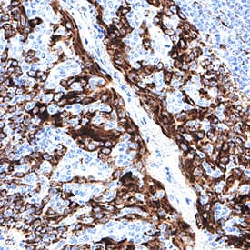
Detection of CKAP4/p63 in Human Tonsil. CKAP4/p63 was detected in immersion fixed paraffin-embedded sections of human tonsil using Mouse Anti-Human CKAP4/p63 Monoclonal Antibody (Catalog # MAB11600) at 5 µg/ml for 1 hour at room temperature followed by incubation with the Anti-Mouse IgG VisUCyte™ HRP Polymer Antibody (Catalog # VC001). Before incubation with the primary antibody, tissue was subjected to heat-induced epitope retrieval using VisUCyte Antigen Retrieval Reagent-Basic (Catalog # VCTS021). Tissue was stained using DAB (brown) and counterstained with hematoxylin (blue). Specific staining was localized to the cell surface and cytoplasm. View our protocol for IHC Staining with VisUCyte HRP Polymer Detection Reagents.
 View Larger
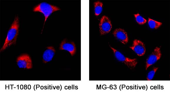
Detection of CKAP4/p63 in HT-1080 and MG‑63 Human Cell Line. CKAP4/p63 was detected in immersion fixed HT-1080 cells and MG‑63 human osteosarcoma cell line using Mouse Anti-Human CKAP4/p63 Monoclonal Antibody (Catalog # MAB11600) at 8 µg/ml for 3 hours at room temperature. Cells were stained using the NorthernLights™ 557-conjugated Anti-Mouse IgG Secondary Antibody (red; Catalog # NL007) and counterstained with DAPI (blue). Specific staining was localized to the endoplasmic reticulum membrane. View our protocol for Fluorescent ICC Staining of Cells on Coverslips.
 View Larger
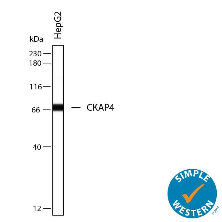
Detection of Human CKAP4/p63 by Simple WesternTM. Simple Western shows lysates of HepG2 human hepatocellular carcinoma cell line, loaded at 0.2 mg/ml. A specific band was detected for CKAP4/p63 at approximately 65 kDa (as indicated) using 4 µg/mL of Mouse Anti-Human CKAP4/p63 Monoclonal Antibody (Catalog # MAB11600). This experiment was conducted under reducing conditions and using the 12‑230 kDa separation system.
Preparation and Storage
- 12 months from date of receipt, -20 to -70 °C as supplied.
- 1 month, 2 to 8 °C under sterile conditions after reconstitution.
- 6 months, -20 to -70 °C under sterile conditions after reconstitution.
Background: CKAP4/p63
CKAP4 (Cytoskeleton-associated protein 4; also CLIMP63 and p63) is a 63-64 kDa molecule that belongs to no known protein family. It is found intracellularly, and on the plasma membrane of select cells such as vascular smooth muscle and Type II Greater alveolar lung cells. CKAP4 has a bimodal distribution. First, it is embedded in the membrane of a compartment that links the ER with the Golgi apparatus. This localization is dependent upon its ability to form homooligomers, and its presence serves to anchor microtubules and direct the formation of tubular ER. Second, it is embedded in the plasma membrane and serves as a receptor for SP-A/surfactant protein-A (in lung) and tPA (in vessels). Human CKAP4 is a 602 amino acid (aa) type II transmembrane nonglycosylated protein. It contains a 106 aa N-terminal cytoplasmic region plus a 475 aa C-terminal luminal domain (aa 128-602). The luminal domain contains three coiled-coil regions (aa 130-214; 256-460; 533-602) plus three utilized phosphorylation sites. CKAP4 undergoes reversible palmitoylation. There are two potential isoform variants. One contains an alternative start at Met269, while another shows a deletion of aa 258-435. Over aa 128-602, human CKAP4 shares 82% aa sequence identity with mouse CKAP4.
Product Datasheets
FAQs
No product specific FAQs exist for this product, however you may
View all Antibody FAQsReviews for Human CKAP4/p63 Antibody
There are currently no reviews for this product. Be the first to review Human CKAP4/p63 Antibody and earn rewards!
Have you used Human CKAP4/p63 Antibody?
Submit a review and receive an Amazon gift card.
$25/€18/£15/$25CAN/¥75 Yuan/¥2500 Yen for a review with an image
$10/€7/£6/$10 CAD/¥70 Yuan/¥1110 Yen for a review without an image
