Human IL-1 beta /IL-1F2 Antibody
Human IL-1 beta /IL-1F2 Antibody Summary
Applications
Human IL-1 beta /IL-1F2 Sandwich Immunoassay
Please Note: Optimal dilutions should be determined by each laboratory for each application. General Protocols are available in the Technical Information section on our website.
Scientific Data
 View Larger
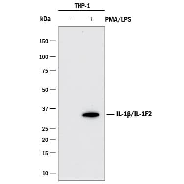
Detection of Human IL‑1 beta /IL‑1F2 by Western Blot. Western blot shows lysates of THP-1 human acute monocytic leukemia cell line untreated (-) or treated (+) with 200 nM PMA for 24 hours and 10 µg/mL LPS for 3 hours. PVDF membrane was probed with 1 µg/mL of Mouse Anti-Human IL-1 beta /IL-1F2 Monoclonal Antibody (Catalog # MAB601R) followed by HRP-conjugated Anti-Mouse IgG Secondary Antibody (Catalog # HAF018). A specific band was detected for IL-1 beta /IL-1F2 at approximately 35 kDa (as indicated). This experiment was conducted under reducing conditions and using Immunoblot Buffer Group 1.
 View Larger
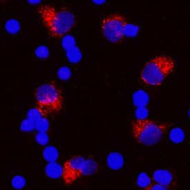
IL‑1 beta /IL‑1F2 in Human PBMCs. IL-1 beta /IL-1F2 was detected in immersion fixed human peripheral blood mononuclear cells (PBMCs) treated with 1 µg/mL LPS and 3 µM monensin for 24 hours using Mouse Anti-Human IL-1 beta /IL-1F2 Monoclonal Antibody (Catalog # MAB601R) at 10 µg/mL for 3 hours at room temperature. Cells were stained using the NorthernLights™ 557-conjugated Anti-Mouse IgG Secondary Antibody (red; Catalog # NL007) and counterstained with DAPI (blue). Specific staining was localized to cytoplasm. View our protocol for Fluorescent ICC Staining of Non-adherent Cells.
 View Larger
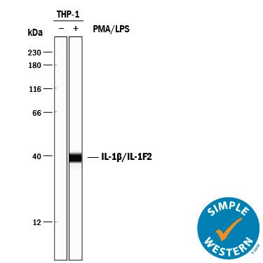
Detection of Human IL‑1 beta /IL‑1F2 by Simple WesternTM. Simple Western lane view shows lysates of THP‑1 human acute monocytic leukemia cell line untreated (-) or treated (+) with 200 nM PMA for 200 nM and 10 μg/mL LPS for 3 hours, loaded at 0.2 mg/mL. A specific band was detected for IL‑1 beta /IL‑1F2 at approximately 37 kDa (as indicated) using 10 µg/mL of Mouse Anti-Human IL‑1 beta /IL‑1F2 Monoclonal Antibody (Catalog # MAB601R). This experiment was conducted under reducing conditions and using the 12-230 kDa separation system.
 View Larger
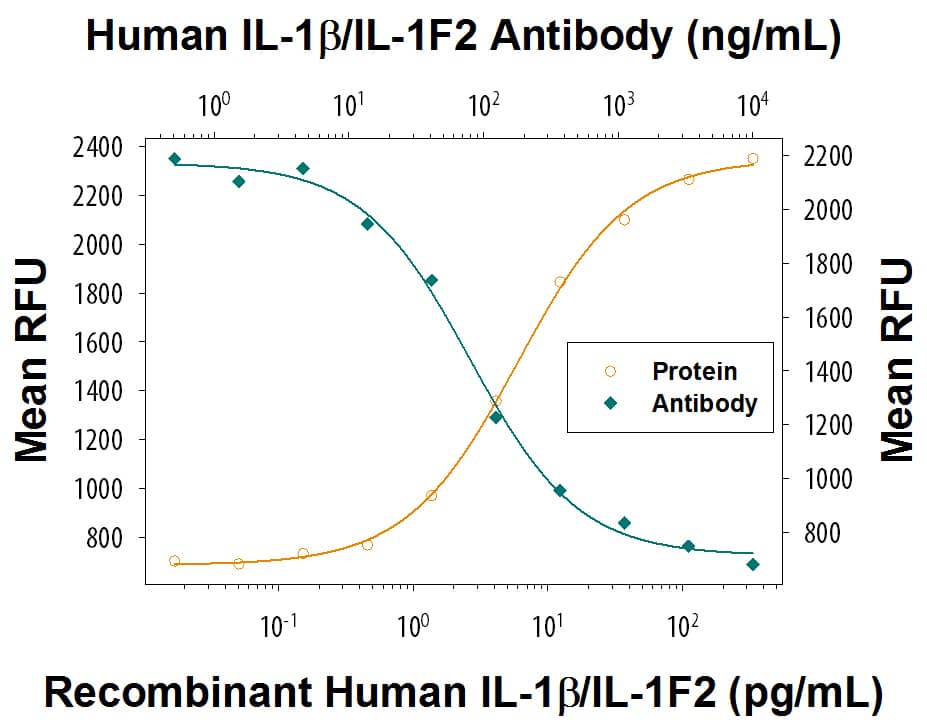
Cell Proliferation Induced by IL-1 beta /IL-1F2 and Neutralization by Human IL-1 beta /IL-1F2 Antibody. Recombinant HumanIL-1 beta /IL-1F2 (Catalog # 201-LB) stimulates proliferation in the the D10.G4.1 mouse helper T cell line in a dose-dependent manner (orange line) as measured by Resazurin (Catalog # AR002). Proliferation elicited by Recombinant Human IL-1 beta /IL-1F2 (50 pg/mL) is neutralized (green line) by increasing concentrations of Mouse Anti-Human IL-1 beta /IL-1F2 Monoclonal Antibody (Catalog # MAB601R). The ND50 is typically 50-200 ng/mL.
Reconstitution Calculator
Preparation and Storage
- 12 months from date of receipt, -20 to -70 °C as supplied.
- 1 month, 2 to 8 °C under sterile conditions after reconstitution.
- 6 months, -20 to -70 °C under sterile conditions after reconstitution.
Background: IL-1 beta/IL-1F2
IL-1 is a name that designates two pleiotropic cytokines, IL-1 alpha (IL-1F1) and IL-1 beta (IL-1F2, IL1B), which are the products of distinct genes. IL-1 alpha and IL-1 beta are structurally related polypeptides that share approximately 21% amino acid (aa) identity in human. Both proteins are produced by a wide variety of cells in response to inflammatory agents, infections, or microbial endotoxins. While IL-1 alpha and IL-1 beta are regulated independently, they bind to the same receptor and exert identical biological effects. IL-1 RI binds directly to IL-1 alpha or IL-1 beta and then associates with IL-1 R accessory protein (IL-1 R3/IL-1 R AcP) to form a high-affinity receptor complex that is competent for signal transduction. IL-1 RII has high affinity for IL-1 beta but functions as a decoy receptor and negative regulator of IL-1 beta activity. IL-1ra functions as a competitive antagonist by preventing IL-1 alpha and IL-1 beta from interacting with IL-1 RI. Intracellular cleavage of the IL-1 beta precursor by Caspase-1/ICE is a key step in the inflammatory response. The 17 kDa molecular weight mature human IL-1 beta shares 96% aa sequence identity with rhesus and 67%-78% with canine, cotton rat, equine, feline, mouse, porcine, and rat IL-1 beta. IL-1 beta functions in a central role in immune and inflammatory responses, bone remodeling, fever, carbohydrate metabolism, and GH/IGF-I physiology. IL-1 beta dysregulation is implicated in many pathological conditions including sepsis, rheumatoid arthritis, inflammatory bowel disease, acute and chronic myelogenous leukemia, insulin-dependent diabetes mellitus, atherosclerosis, neuronal injury, and aging-related diseases.
Product Datasheets
FAQs
No product specific FAQs exist for this product, however you may
View all Antibody FAQsReviews for Human IL-1 beta /IL-1F2 Antibody
There are currently no reviews for this product. Be the first to review Human IL-1 beta /IL-1F2 Antibody and earn rewards!
Have you used Human IL-1 beta /IL-1F2 Antibody?
Submit a review and receive an Amazon gift card.
$25/€18/£15/$25CAN/¥75 Yuan/¥2500 Yen for a review with an image
$10/€7/£6/$10 CAD/¥70 Yuan/¥1110 Yen for a review without an image


