Human Mesothelin Antibody Summary
Glu296-Gly580
Accession # Q13421
Applications
Please Note: Optimal dilutions should be determined by each laboratory for each application. General Protocols are available in the Technical Information section on our website.
Scientific Data
 View Larger
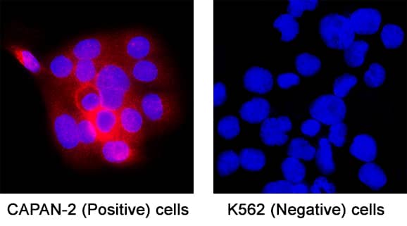
Detection of Mesothelin in Capan-2 (positive) and K562 cells (negative). Mesothelin was detected in immersion fixed Capan‑2 human pancreatic adenocarcinoma cells (positive), and absent in K562 human chronic myelogenous leukemia cells (negative) using Mouse Anti-Human Mesothelin Monoclonal Antibody (Catalog # MAB11417) at 8 µg/mL for 3 hours at room temperature. Cells were stained using the NorthernLights™ 557-conjugated Anti-Mouse IgG Secondary Antibody (red; Catalog # NL007) and counterstained with DAPI (blue). Specific staining was localized to cell cytoplasm. View our protocol for Fluorescent ICC Staining of Cells on Coverslips.
Reconstitution Calculator
Preparation and Storage
- 12 months from date of receipt, -20 to -70 °C as supplied.
- 1 month, 2 to 8 °C under sterile conditions after reconstitution.
- 6 months, -20 to -70 °C under sterile conditions after reconstitution.
Background: Mesothelin
Mesothelin, also known as CAK1 and ERC, is derived from a 70 kDa precursor that also includes Megakaryocyte Potentiating Factor (MPF) (1-3). The 70 kDa precursor is expressed on the cell surface where it is cleaved at a dibasic proteolytic site to release the 32 kDa glycosylated MPF (3, 4). MPF is a cytokine that potentiates IL-3 induced megakaryocyte colony formation (2, 5). The term Mesothelin refers to the 40 kDa glycosylated protein which remains attached to the cell surface via a GPI linkage. Alternate splicing generates additional Mesothelin isoforms that have either an eight amino acid insertion following Ser408 or a substituted C‑terminal region with no GPI anchor (6). This recombinant human Mesothelin lacks the 8 aa insertion, and within aa 296-580 it shares 59% sequence identity with mouse and rat Mesothelin. Mesothelin is normally expressed on mesothelial cells in the pleura, pericardium, and peritoneum as well as in the developing and postnatal pancreas (1, 7). It is up‑regulated in mesotheliomas and a range of carcinomas and adenomas (8 ‑ 11). Mesothelin promotes tumor cell proliferation, migration, anchorage-independent growth, and tumor progression (10, 12). It is coexpressed with the tumor antigen CA125/MUC16 on advanced ovarian adenocarcinomas and interacts with this molecule to support cell adhesion (13). A soluble form of Mesothelin is released from tumor cells into the serum or tissue effusions (11, 14, 15).
- Hassan, R. et al. (2004) Clin. Cancer Res. 10:3937.
- Kojima, T. et al. (1995) J. Biol. Chem. 270:21984.
- Chang, K. and I. Pastan (1996) Proc. Natl. Acad. Sci. 93:136.
- Onda, M. et al. (2006) Clin. Cancer Res. 12:4225.
- Yamaguchi, N. et al. (1994) J. Biol. Chem. 269:805.
- Muminova, Z.E. et al. (2004) BMC Cancer 4:19.
- Hou, L.-Q. et al. (2008) Develop. Growth Differ. 50:531.
- Ordonez, N.G. (2003) Mod. Pathol. 16:192.
- Argani, P. et al. (2001) Clin. Cancer Res. 7:3862.
- Li, M. et al. (2008) Mol. Cancer Ther. 7:286.
- Scholler, N. et al. (1999) Proc. Natl. Acad. Sci. 96:11531.
- Uehara, N. et al. (2008) Mol. Cancer Res. 6:186.
- Rump, A. et al. (2004) J. Biol. Chem. 279:9190.
- Ho, M. and M.O. Lively (2006) Cancer Epidemiol. Biomarkers Prev. 15:1751.
- Robinson, B.W.S. et al. (2003) Lancet 362:1612.
Product Datasheets
FAQs
No product specific FAQs exist for this product, however you may
View all Antibody FAQsReviews for Human Mesothelin Antibody
There are currently no reviews for this product. Be the first to review Human Mesothelin Antibody and earn rewards!
Have you used Human Mesothelin Antibody?
Submit a review and receive an Amazon gift card.
$25/€18/£15/$25CAN/¥75 Yuan/¥2500 Yen for a review with an image
$10/€7/£6/$10 CAD/¥70 Yuan/¥1110 Yen for a review without an image

