Human Phospho-p53 (S15) Antibody
Human Phospho-p53 (S15) Antibody Summary
Accession # P04637
Applications
Please Note: Optimal dilutions should be determined by each laboratory for each application. General Protocols are available in the Technical Information section on our website.
Scientific Data
 View Larger
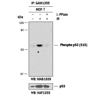
Detection of Human Phospho-p53 (S15) by Western Blot. Western blot shows p53 immunoprecipitated from lysates of MCF-7 human breast cancer cell line using Human/Mouse/Rat p53 Agarose-conjugated Antigen Affinity-purified Polyclonal Antibody (Catalog # GAF1355). MCF-7 cell line was untreated (-) or exposed (+) to 10 Gy ionizing radiation (IR) for 3 hours. PVDF membrane was probed with 1-2 µg/mL Mouse Anti-Human Phospho-p53 (S15) Monoclonal Antibody (Catalog # MAB1839) followed by HRP-conjugated Anti-Mouse IgG Secondary Antibody (Catalog # HAF007). A specific band for Phospho-p53 (S15) was detected at approximately 53 kDa (as indicated). The phospho-specificity of this antibody was supported by decreased labeling following treatment with 600 U ?-phosphatase (?-PPase) for 1 hour. For additional reference the membrane was stripped and reprobed with 1:5000 dilution Human/Mouse/Rat p53 HRP-conjugated Antigen Affinity-purified Polyclonal Antibody HAF1355 (lower panel, Catalog # HAF1355) This experiment was conducted under reducing conditions and using Immunoblot Buffer Group 1.
 View Larger
Detection of p53 in camptothecin-treated MCF‑7 Human Cell Line by Flow Cytometry. MCF-7 human breast cancer cell line was unstimulated (light orange open histogram) or treated with 1 µM camphtothecin for 6 hours (dark orange filled histogram), then stained with Mouse Anti-Human Phospho-p53 (S15) Monoclonal Antibody (Catalog # MAB1839) or isotype control (Catalog # MAB002, blue open histogram), followed by Phycoerythrin-conjugated Anti-Mouse IgG F(ab')2Secondary Antibody (Catalog # F0102B). To facilitate intracellular staining, cells were fixed with paraformaldehyde and permeabilized with saponin.
Reconstitution Calculator
Preparation and Storage
- 12 months from date of receipt, -20 to -70 °C as supplied.
- 1 month, 2 to 8 °C under sterile conditions after reconstitution.
- 6 months, -20 to -70 °C under sterile conditions after reconstitution.
Background: p53
The p53 tumor suppressor protein acts to enforce cell cycle checkpoints or signal apoptosis in cells that have incurred genotoxic damage. The ATM or ATR kinases can phosphorylate p53 at serine 15 (S15), which leads to cell cycle arrest. Serine 15 phosphorylation leads to p53 stabilization and enhances transactivation of p53 target genes.
Product Datasheets
Citation for Human Phospho-p53 (S15) Antibody
R&D Systems personnel manually curate a database that contains references using R&D Systems products. The data collected includes not only links to publications in PubMed, but also provides information about sample types, species, and experimental conditions.
1 Citation: Showing 1 - 1
-
OMIP-045: Characterizing human head and neck tumors and cancer cell lines with mass cytometry
Authors: TM Brodie, V Tosevski, M Medová
Cytometry A, 2018-04-12;0(0):.
Species: Human
Sample Types: Whole Cells
Applications: Flow Cytometry
FAQs
No product specific FAQs exist for this product, however you may
View all Antibody FAQsReviews for Human Phospho-p53 (S15) Antibody
There are currently no reviews for this product. Be the first to review Human Phospho-p53 (S15) Antibody and earn rewards!
Have you used Human Phospho-p53 (S15) Antibody?
Submit a review and receive an Amazon gift card.
$25/€18/£15/$25CAN/¥75 Yuan/¥2500 Yen for a review with an image
$10/€7/£6/$10 CAD/¥70 Yuan/¥1110 Yen for a review without an image


