Human TIMP-1 Antibody Summary
Cys24-Ala207
Accession # Q6FGX5
Applications
Please Note: Optimal dilutions should be determined by each laboratory for each application. General Protocols are available in the Technical Information section on our website.
Scientific Data
 View Larger
Detection of Human TIMP‑1 by Western Blot. Western blot shows lysates of human lung tissue and human prostate tissue. PVDF membrane was probed with 1 µg/mL of Goat Anti-Human TIMP-1 Antigen Affinity-purified Polyclonal Antibody (Catalog # AF970) followed by HRP-conjugated Anti-Goat IgG Secondary Antibody (Catalog # HAF017). A specific band was detected for TIMP-1 at approximately 25 kDa (as indicated). This experiment was conducted under reducing conditions and using Immunoblot Buffer Group 1.
 View Larger
Detection of Human TIMP‑1 by Western Blot. Western blot shows lysates of PC-3 human prostate cancer cell line, HT-29 human colon adenocarcinoma cell line, and SK-OV-3 human ovarian adenocarcinoma cell line. PVDF membrane was probed with 5 µg/mL of Goat Anti-Human TIMP-1 Antigen Affinity-purified Polyclonal Antibody (Catalog # AF970) followed by HRP-conjugated Anti-Goat IgG Secondary Antibody (Catalog # HAF017). A specific band was detected for TIMP-1 at approximately 26 kDa (as indicated). This experiment was conducted under reducing conditions and using Immunoblot Buffer Group 1.
 View Larger
TIMP‑1 in Human Colon Cancer Tissue. TIMP-1 was detected in immersion fixed paraffin-embedded sections of human colon cancer tissue using Goat Anti-Human TIMP-1 Antigen Affinity-purified Polyclonal Antibody (Catalog # AF970) at 1 µg/mL for 1 hour at room temperature followed by incubation with the Anti-Goat IgG VisUCyte™ HRP Polymer Antibody (Catalog # VC004). Before incubation with the primary antibody, tissue was subjected to heat-induced epitope retrieval using Antigen Retrieval Reagent-Basic (Catalog # CTS013). Tissue was stained using DAB (brown) and counterstained with hematoxylin (blue). Specific staining was localized to cytoplasm and extracellular space. View our protocol for IHC Staining with VisUCyte HRP Polymer Detection Reagents.
 View Larger
Detection of Human TIMP‑1 by Simple WesternTM. Simple Western lane view shows lysates of PC-3 human prostate cancer cell line, HT-29 human colon adenocarcinoma cell line, and human lung tissue, loaded at 0.2 mg/mL. A specific band was detected for TIMP-1 at approximately 42-45 kDa (as indicated) using 50 µg/mL of Goat Anti-Human TIMP-1 Antigen Affinity-purified Polyclonal Antibody (Catalog # AF970) followed by 1:50 dilution of HRP-conjugated Anti-Goat IgG Secondary Antibody (Catalog # HAF109). This experiment was conducted under reducing conditions and using the 12-230 kDa separation system.
 View Larger
Western Blot Shows Human TIMP‑1 Specificity by Using Knockout Cell Line. Western blot shows lysates of SK-OV-3 human ovarian adenocarcinoma cell line and TIMP-1 knockout SK-OV-3 cell line (KO). PVDF membrane was probed with 2 µg/mL of Goat Anti-Human TIMP-1 Antigen Affinity-purified Polyclonal Antibody (Catalog # AF970) followed by HRP-conjugated Anti-Goat IgG Secondary Antibody (Catalog # HAF017). A specific band was detected for TIMP-1 at approximately 25 kDa (as indicated) in the parental SK-OV-3 cell line, but is not detectable in knockout SK-OV-3 cell line. GAPDH (Catalog # AF5718) is shown as a loading control. This experiment was conducted under reducing conditions and using Immunoblot Buffer Group 1.
 View Larger
Neutralization of TIMP‑1 Activity by Human TIMP‑1 Antibody. Recombinant Human MMP-2 (0.2 µg/mL, Catalog # 902-MP) activity is measured in the presence of Recombinant Human TIMP-1 (0.1 µg/mL, Catalog # 970-TM) that has been preincubated with increasing concentrations of Goat Anti-Human TIMP-1 Antigen Affinity-purified Polyclonal Antibody (Catalog # AF970). The ND50 is typically 1 µg/mL.
 View Larger
Detection of Human TIMP-1 by Immunohistochemistry-Paraffin IHC staining result investigating TR4 level and macrophage infiltration in tumor tissues of PCa patients, as well as TIMP-1/MMP2/MMP9 signaling. IHC staining was performed using TR4 antibody (1:300),CD68 antibody (1:200),TIMP-1 antibody (1:200),MMP2 antibody (1:200),MMP9 antibody (1:200). Image collected and cropped by CiteAb from the following open publication (https://pubmed.ncbi.nlm.nih.gov/25623427), licensed under a CC-BY license. Not internally tested by R&D Systems.
 View Larger
Detection of Human TIMP-1 by Proximity Ligation Assay Univariate survival analysis of disease free survival (DFS) in plasma for all 465 patients. A) MMP-9:TIMP-1 measured by ELISA. B) MMP-9:TIMP-1 measured by PLA. Patients are divided into four groups of equal size (Q1-Q4) according to increasing plasma MMP-9:TIMP-1 levels; Q1 being the group with the lowest level. Image collected and cropped by CiteAb from the following open publication (https://bmccancer.biomedcentral.com/articles/10.1186/1471-2407-13-598), licensed under a CC-BY license. Not internally tested by R&D Systems.
Reconstitution Calculator
Preparation and Storage
- 12 months from date of receipt, -20 to -70 °C as supplied.
- 1 month, 2 to 8 °C under sterile conditions after reconstitution.
- 6 months, -20 to -70 °C under sterile conditions after reconstitution.
Background: TIMP-1
Tissue inhibitors of metalloproteinases or TIMPs are a family of proteins that regulate the activation and proteolytic activity of the zinc enzymes known as matrix metalloproteinases (MMPs). There are four members of the family, TIMP-1, TIMP-2, TIMP-3 and TIMP-4. TIMP-1 is a glycoprotein with a molecular mass of 28 kDa produced by a wide range of cell types. TIMP-1 inhibits active MMP-mediated proteolysis by forming an N-terminal, non-covalent binary complex with the MMP active site. TIMP-1 also associates C-terminally with Pro-MMP-9 in a complex which may play a role in regulating activation. Independent of MMPs, TIMP-1 has been shown to have a role in tissue homeostasis.
Product Datasheets
Citations for Human TIMP-1 Antibody
R&D Systems personnel manually curate a database that contains references using R&D Systems products. The data collected includes not only links to publications in PubMed, but also provides information about sample types, species, and experimental conditions.
23
Citations: Showing 1 - 10
Filter your results:
Filter by:
-
CD49f Is a Novel Marker of Functional and Reactive Human iPSC-Derived Astrocytes
Authors: Barbar L, Jain T, Zimmer M et al.
Neuron
-
The periosteum provides a stromal defence against cancer invasion into the bone
Authors: Nakamura, K;Tsukasaki, M;Tsunematsu, T;Yan, M;Ando, Y;Huynh, NC;Hashimoto, K;Gou, Q;Muro, R;Itabashi, A;Iguchi, T;Okamoto, K;Nakamura, T;Nakano, K;Okamura, T;Ueno, T;Ito, K;Ishimaru, N;Hoshi, K;Takayanagi, H;
Nature
Species: Mouse, Transgenic Mouse
Sample Types: Whole Tissue
Applications: Immunohistochemistry -
Myeloma Microenvironmental TIMP1 Induces the Invasive Phenotype in Fibroblasts to Modulate Disease Progression
Authors: R Ishihara, T Oda, Y Murakami, I Matsumura, S Watanabe, Y Asao, Y Masuda, N Gotoh, T Kasamatsu, H Takei, N Kobayashi, N Sasaki, T Saitoh, H Murakami, H Handa
International Journal of Molecular Sciences, 2023-01-22;24(3):.
Species: Human
Sample Types: Whole Cells
Applications: Neutralization -
Inhibition of Monoacylglycerol Lipase Decreases Angiogenic Features of Endothelial Cells via Release of Tissue Inhibitor of Metalloproteinase-1 from Lung Cancer Cells
Authors: Wittig, F;Henkel, L;Prüser, J;Merkord, J;Ramer, R;Hinz, B;
Cells
Species: Human
Sample Types: Whole Tissue
Applications: Immunohistochemistry -
Improved contractile potential in detrusor microtissues from pediatric patients with end stage lower urinary tract dysfunction
Authors: Gerwinn T, Salemi S, Schori LJ et al.
Frontiers in Cell and Developmental Biology
-
Single cell transcriptomic landscape of diabetic foot ulcers
Authors: G Theocharid, BE Thomas, D Sarkar, HL Mumme, WJR Pilcher, B Dwivedi, T Sandoval-S, RF Sîrbulescu, A Kafanas, I Mezghani, P Wang, A Lobao, IS Vlachos, B Dash, HC Hsia, V Horsley, SS Bhasin, A Veves, M Bhasin
Nature Communications, 2022-01-10;13(1):181.
Species: Human
Sample Types: Whole Tissue
Applications: IHC -
Inhibition of epithelial-mesenchymal transition in retinal pigment epithelial cells by a retinoic acid receptor-&alpha agonist
Authors: Y Kobayashi, K Tokuda, C Yamashiro, F Higashijim, T Yoshimoto, M Ota, T Ogata, A Ashimori, M Hatano, M Kobayashi, SH Uchi, M Wakuta, K Kimura
Scientific Reports, 2021-06-04;11(1):11842.
Species: Human
Sample Types: Cell Lysates
Applications: Western Blot -
Heroin Seeking and Extinction From Seeking Activate Matrix Metalloproteinases at Synapses on Distinct Subpopulations of Accumbens Cells
Authors: Vivian C. Chioma, Anna Kruyer, Ana-Clara Bobadilla, Ariana Angelis, Zachary Ellison, Ritchy Hodebourg et al.
Biological Psychiatry
-
IL35 predicts prognosis in gastric cancer and is associated with angiogenesis by altering TIMP1, PAI1, and IGFBP1
Authors: X Li, N Niu, J Sun, Y Mou, X He, L Mei
FEBS Open Bio, 2020-11-09;0(0):.
Species: Human
Sample Types: Whole Cells
Applications: Neutralization -
The Metalloproteinase ADAMTS5 Is Expressed by Interstitial Inflammatory Cells in IgA Nephropathy and Is Proteolytically Active on the Kidney Matrix
Authors: S Taylor, M Whitfield, J Barratt, A Didangelos
J. Immunol., 2020-09-11;0(0):.
Species: Human
Sample Types: Whole Tissue
Applications: IHC -
Circulating Tumor Cells Characterization Revealed TIMP1 as a Potential Therapeutic Target in Ovarian Cancer
Authors: M Abreu, P Cabezas-Sa, L Alonso-Alc, A Ferreirós, P Mondelo-Ma, RM Lago-Lestó, A Abalo, E Díaz, S Palacios-Z, A Rojo-Sebas, R López-Lópe, L Sánchez, G Moreno-Bue, L Muinelo-Ro
Cells, 2020-05-14;9(5):.
Species: Human
Sample Types: Whole Cell
Applications: Western Blot -
Looking for a Better Characterization of Triple-Negative Breast Cancer by Means of Circulating Tumor Cells
Authors: M Abreu, P Cabezas-Sa, T Pereira-Ve, C Falo, A Abalo, I Morilla, T Curiel, J Cueva, C Rodríguez, V Varela-Pos, R Lago-Lestó, P Mondelo, P Palacios, G Moreno-Bue, A Cano, T García-Cab, MÁ Pujana, L Sánchez-Pi, C Costa, R López, L Muinelo-Ro
J Clin Med, 2020-01-27;9(2):.
Species: Human
Sample Types: Cell Culture Lysates
Applications: Western Blot -
SILAC Analysis Reveals Increased Secretion of Hemostasis-Related Factors by Senescent Cells
Authors: CD Wiley, S Liu, C Limbad, AM Zawadzka, J Beck, M Demaria, R Artwood, F Alimirah, JA Lopez-Domi, C Kuehnemann, SR Danielson, N Basisty, HG Kasler, TR Oron, PY Desprez, SD Mooney, BW Gibson, B Schilling, J Campisi, P Kapahi
Cell Rep, 2019-09-24;28(13):3329-3337.e5.
Species: Human
Sample Types: Cell Culture Supernates
Applications: Western Blot -
Higher levels of TIMP-1 expression are associated with a poor prognosis in triple-negative breast cancer
Authors: Guangcun Cheng, Xuemei Fan, Mingang Hao, Jinglong Wang, Xiaoming Zhou, Xueqing Sun
Molecular Cancer
-
Targeting TR4 nuclear receptor suppresses prostate cancer invasion via reduction of infiltrating macrophages with alteration of the TIMP-1/MMP2/MMP9 signals.
Authors: Ding X, Yang D, Xia L, Chen B, Yu S, Niu Y, Wang M, Li G, Chang C
Mol Cancer, 2015-01-27;14(0):16.
Species: Human
Sample Types: Whole Tissue
Applications: IHC-P -
Plasma levels of the MMP-9:TIMP-1 complex as prognostic biomarker in breast cancer: a retrospective study.
Authors: Thorsen S, Christensen S, Wurtz S, Lundberg M, Nielsen B, Vinther L, Knowles M, Gee N, Fredriksson S, Moller S, Brunner N, Schrohl A, Stenvang J
BMC Cancer, 2013-12-13;13(0):598.
Species: Human
Sample Types: Plasma
Applications: Proximity Ligation Assay (PLA) -
Adipocytes promote ovarian cancer metastasis and provide energy for rapid tumor growth.
Authors: Nieman KM, Kenny HA, Penicka CV, Ladanyi A, Buell-Gutbrod R, Zillhardt MR, Romero IL, Carey MS, Mills GB, Hotamisligil GS, Yamada SD, Peter ME, Gwin K, Lengyel E
Nat. Med., 2011-10-30;17(11):1498-503.
Species: Human
Sample Types: In Vivo
Applications: In Vivo -
Breast cancer cells induce cancer-associated fibroblasts to secrete hepatocyte growth factor to enhance breast tumorigenesis.
Authors: Tyan SW, Kuo WH, Huang CK
PLoS ONE, 2011-01-13;6(1):e15313.
Species: Human
Sample Types: Whole Cells
Applications: Neutralization -
Primary human acute myelogenous leukemia cells release matrix metalloproteases and their inhibitors: release profile and pharmacological modulation.
Authors: Reikvam H, Hatfield KJ, Oyan AM, Kalland KH, Kittang AO, Bruserud O
Eur. J. Haematol., 2009-11-17;84(3):239-51.
Species: Human
Sample Types: Whole Cells
Applications: Neutralization -
Fibroblast-conditioned media promote human sarcoma cell invasion.
Authors: Bittner JG, Wilson M, Shah MB, Albo D, Feig BW, Wang TN
Surgery, 2008-09-14;145(1):42-7.
Species: Human
Sample Types: Cell Lysates
Applications: Western Blot -
Coregulation of vascular tube stabilization by endothelial cell TIMP-2 and pericyte TIMP-3.
Authors: Saunders WB, Bohnsack BL, Faske JB, Anthis NJ, Bayless KJ, Hirschi KK, Davis GE
J. Cell Biol., 2006-10-09;175(1):179-91.
Species: Human
Sample Types: Cell Lysates
Applications: Western Blot -
Median raphe serotonergic neurons projecting to the interpeduncular nucleus control preference and aversion
Authors: H Kawai, Y Bouchekiou, N Nishitani, K Niitani, S Izumi, H Morishita, C Andoh, Y Nagai, M Koda, M Hagiwara, K Toda, H Shirakawa, K Nagayasu, Y Ohmura, M Kondo, K Kaneda, M Yoshioka, S Kaneko
Nature Communications, 2022-12-22;13(1):7708.
-
Involvement of TIMP-1 in PECAM-1-mediated tumor dissemination
Authors: Abraham V, Cao G, Parambath A et al.
Int J Onc
FAQs
No product specific FAQs exist for this product, however you may
View all Antibody FAQsReviews for Human TIMP-1 Antibody
Average Rating: 4.5 (Based on 2 Reviews)
Have you used Human TIMP-1 Antibody?
Submit a review and receive an Amazon gift card.
$25/€18/£15/$25CAN/¥75 Yuan/¥2500 Yen for a review with an image
$10/€7/£6/$10 CAD/¥70 Yuan/¥1110 Yen for a review without an image
Filter by:
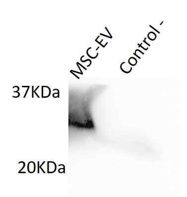
Negative control: murine macrophages.
MSC-EV: mesenchymal stem cells derived extracellular vesicles


