Mouse EGF Quantikine ELISA Kit Summary
Product Summary
Precision
Cell Culture Supernates, Tissue Homogenates, Urine
| Intra-Assay Precision | Inter-Assay Precision | |||||
|---|---|---|---|---|---|---|
| Sample | 1 | 2 | 3 | 1 | 2 | 3 |
| n | 20 | 20 | 20 | 27 | 28 | 27 |
| Mean (pg/mL) | 35.4 | 78 | 244 | 28 | 65.3 | 232 |
| Standard Deviation | 3.2 | 7.4 | 11.8 | 2.6 | 4.6 | 18.4 |
| CV% | 9 | 9.5 | 4.8 | 9.3 | 7 | 7.9 |
Serum, EDTA Plasma, Heparin Plasma
| Intra-Assay Precision | Inter-Assay Precision | |||||
|---|---|---|---|---|---|---|
| Sample | 1 | 2 | 3 | 1 | 2 | 3 |
| n | 20 | 20 | 20 | 24 | 25 | 24 |
| Mean (pg/mL) | 34.6 | 86.6 | 304 | 34.7 | 82.8 | 299 |
| Standard Deviation | 3.4 | 7.9 | 14.7 | 3.2 | 7.7 | 28.4 |
| CV% | 9.8 | 9.1 | 4.8 | 9.2 | 9.3 | 9.5 |
Recovery
The recovery of mouse EGF spiked to three levels throughout the range of the assay in various matrices was evaluated.
| Sample Type | Average % Recovery | Range % |
|---|---|---|
| Cell Culture Supernates (n=4) | 109 | 101-119 |
| EDTA Plasma (n=4) | 97 | 87-106 |
| Heparin Plasma (n=4) | 95 | 87-105 |
| Serum (n=4) | 96 | 83-107 |
| Tissue Homogenates (n=3) | 93 | 89-101 |
Linearity
Scientific Data
 View Larger
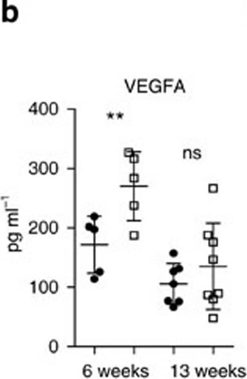
Detection of Mouse EGF by ELISA STAT3 alters tumour microenvironment and angiogenesis.(a) IHC analysis of von Willebrand Factor (vWF) and CD31 is shown in consecutive sections of murine lung tumours of the indicated genotype. On right, CD31+ counts per tumour area (mm2) quantification at indicated time points. Data were analysed by Student’s t-test and displayed as mean±s.e.m. n≥5 tumours per mouse with n≥4 mice per genotype and time point. Scale bar, 50 μm. (b) ELISA of VEGFA levels in lung tumour lysates at indicated time points. Data were analysed by Student’s t-test and displayed as mean±s.d., n≥6 mice per genotype and time point. (c) Expression levels of Pdgfa in total lungs were measured by quantitative real-time PCR at indicated time points. Values are presented as fold change of relative mRNA expression compared with Stat3 delta Lep/ delta Lep:Kras+/+ mice. Data were analysed by one-way analysis of variance (ANOVA) with Tukey’s multiple comparison test and are shown as mean± s.d., n≥5 animals/genotype. (d) Flow cytometric analysis of Cd11b+Gr1+ granulocytes and Cd11b+ F4/80+ macrophages in bronchoalveolar lavage (BAL) at 6 and 13 weeks post AdCre. Data were analysed by one-way ANOVA with Tukey’s multiple comparison test and are shown as mean±s.e.m. (e) Flow cytometric analysis of myeloid-derived suppressor cell (MDSC) subsets at 6 and 13 weeks post AdCre. Data were analysed by one-way ANOVA with Tukey’s multiple comparison test and are shown as mean± s.e.m. (f) Ratio of CD4+/CD8+ T-cell counts in BAL are shown. Data were analysed by Kruskal–Wallis test with Dunn’s multiple comparison testing and are shown as mean±s.e.m. Data displayed in d–f are n≥6 mice per genotype and time point, 13-week group represents two independent experiments. (g) Flow cytometric analysis of Cd11b+Gr1+ and Cd11b+ F4/80+ cells of A549 shControl versus A549-shSTAT3 xenograft tumours. Data were analysed by Student’s t-test and are displayed as mean±s.e.m. (n≥8 tumours; ≥4 mice per group). (h) IHC analysis and representative pictures of CD31+ counts per xenograft tumour area (mm2). Scale bar, 100 μm (n=8 tumours/≥4 mice). Data were analysed by Student’s t-test and are shown as mean±s.e.m. For all graphs: *P<0.05; **P<0.01; ***P<0.001. Image collected and cropped by CiteAb from the following publication (https://www.nature.com/articles/ncomms7285), licensed under a CC-BY license. Not internally tested by R&D Systems.
Product Datasheets
Preparation and Storage
Background: EGF
EGF (Epidermal Growth Factor) is expressed as a transmembrane protein that is proteolytically cleaved to generate soluble forms. It signals through EGF R/ErbB1 which heterodimerizes with ErbB2, ErbB3, or ErbB4 and can also associate with PDGF R beta and HGF R/c-MET. During development, EGF regulates thymocyte differentiation, neuroglia production, and adipocyte maturation. In the adult, EGF plays a role in mammary gland lactogenesis, fibroblast mitosis, dissociation of the extracellular matrix, and cell migration.
Assay Procedure
Refer to the product- Prepare all reagents, standard dilutions, and samples as directed in the product insert.
- Remove excess microplate strips from the plate frame, return them to the foil pouch containing the desiccant pack, and reseal.
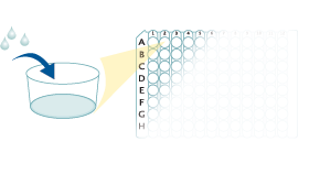
- Add 50 µL of Assay Diluent to each well.
- Add 50 µL of Standard, Control, or sample to each well. Cover with a plate sealer, and incubate at room temperature for 2 hours.
- Aspirate each well and wash, repeating the process 4 times for a total of 5 washes.
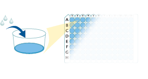
- Add 100 µL of Conjugate to each well. Cover with a new plate sealer, and incubate at room temperature for 2 hours.
- Aspirate and wash 5 times.
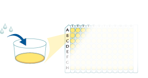
- Add 100 µL Substrate Solution to each well. Incubate at room temperature for 30 minutes. PROTECT FROM LIGHT.
- Add 100 µL of Stop Solution to each well. Read at 450 nm within 30 minutes. Set wavelength correction to 540 nm or 570 nm.


Citations for Mouse EGF Quantikine ELISA Kit
R&D Systems personnel manually curate a database that contains references using R&D Systems products. The data collected includes not only links to publications in PubMed, but also provides information about sample types, species, and experimental conditions.
14
Citations: Showing 1 - 10
Filter your results:
Filter by:
-
Optogenetic Stimulation of the Cardiac Vagus Nerve to Promote Heart Regenerative Repair after Myocardial Infarction
Authors: Han, Y;Wei, X;Chen, G;Shao, E;Zhou, Y;Li, Y;Xiao, Z;Shi, X;Zheng, H;Huang, S;Chen, Y;Wang, Y;Zhang, Y;Liao, Y;Liao, W;Bin, J;Wang, Y;Li, X;
International journal of biological sciences
Species: Mouse
Sample Types: Cell Culture Supernates
-
Enhanced cellular engraftment of adipose-derived mesenchymal stem cell spheroids by using nanosheets as scaffolds
Authors: H Nagano, Y Suematsu, M Takuma, S Aoki, A Satoh, E Takayama, M Kinoshita, Y Morimoto, S Takeoka, T Fujie, T Kiyosawa
Scientific Reports, 2021-07-14;11(1):14500.
Species: Mouse
Sample Types: Tissue Homogenates
-
Probiotic-Derived Polyphosphate Accelerates Intestinal Epithelia Wound Healing through Inducing Platelet-Derived Mediators
Authors: S Isozaki, H Konishi, M Fujiya, H Tanaka, Y Murakami, S Kashima, K Ando, N Ueno, K Moriichi, T Okumura
Mediators of Inflammation, 2021-03-29;2021(0):5582943.
Species: Rat
Sample Types: Plasma
-
Adipose-derived mesenchymal stem cells regenerate radioiodine-induced salivary gland damage in a murine model
Authors: JW Kim, JM Kim, ME Choi, SK Kim, YM Kim, JS Choi
Sci Rep, 2019-10-31;9(1):15752.
Species: Mouse
Sample Types: Saliva
-
Autophagy regulates the therapeutic potential of adipose-derived stem cells in LPS-induced pulmonary microvascular barrier damage
Authors: C Li, J Pan, L Ye, H Xu, B Wang, H Xu, L Xu, T Hou, D Zhang
Cell Death Dis, 2019-10-23;10(11):804.
Species: Mouse
Sample Types: Cell Culture Supernates
-
Attached stratified mucus separates bacteria from the epithelial cells in COPD lungs
Authors: JA Fernández-, D Fakih, L Arike, AM Rodríguez-, B Martínez-A, E Skansebo, S Jackson, J Root, D Singh, C McCrae, CM Evans, A Åstrand, A Ermund, GC Hansson
JCI Insight, 2018-09-06;3(17):.
Species: Mouse
Sample Types: BALF
-
Decellularized Diaphragmatic Muscle Drives a Constructive Angiogenic Response In Vivo
Authors: ME Alvarèz Fa, M Piccoli, C Franzin, A Sgrò, A Dedja, L Urbani, E Bertin, C Trevisan, P Gamba, AJ Burns, P De Coppi, M Pozzobon
Int J Mol Sci, 2018-04-28;19(5):.
Species: Mouse
Sample Types: Tissue Homogenates
-
Corticotrophin-releasing hormone and corticosterone impair development of preimplantation embryos by inducing oviductal cell apoptosis via activating the Fas system: an in vitro study
Authors: XW Tan, CL Ji, LL Zheng, J Zhang, HJ Yuan, S Gong, J Zhu, JH Tan
Hum. Reprod., 2017-08-01;0(0):1-15.
Species: Mouse
Sample Types: Cell Culture Supernates
-
Radioprotective effects of Keratinocyte Growth Factor-1 against irradiation-induced salivary gland hypofunction
Authors: JS Choi, HS Shin, HY An, YM Kim, JY Lim
Oncotarget, 2017-02-21;8(8):13496-13508.
Species: Mouse
Sample Types: Saliva
-
Controlled water vapor transmission rate promotes wound-healing via wound re-epithelialization and contraction enhancement
Authors: R Xu, H Xia, W He, Z Li, J Zhao, B Liu, Y Wang, Q Lei, Y Kong, Y Bai, Z Yao, R Yan, H Li, R Zhan, S Yang, G Luo, J Wu
Sci Rep, 2016-04-18;6(0):24596.
Species: Mouse
Sample Types: Whole Tissue
-
Intranasal epidermal growth factor treatment rescues neonatal brain injury.
Authors: Scafidi J, Hammond T, Scafidi S, Ritter J, Jablonska B, Roncal M, Szigeti-Buck K, Coman D, Huang Y, McCarter R, Hyder F, Horvath T, Gallo V
Nature, 2013-12-25;506(7487):230-4.
Species: Mouse
Sample Types: Tissue Homogenates
-
Mesenchymal stromal cells improve salivary function and reduce lymphocytic infiltrates in mice with Sjogren's-like disease.
Authors: Khalili S, Liu Y, Kornete M, Roescher N, Kodama S, Peterson A, Piccirillo CA, Tran SD
PLoS ONE, 2012-06-07;7(6):e38615.
Species: Mouse
Sample Types: Saliva
-
Calpain mediates pulmonary vascular remodeling in rodent models of pulmonary hypertension, and its inhibition attenuates pathologic features of disease.
Authors: Ma W, Han W, Greer PA, Tuder RM, Toque HA, Wang KK, Caldwell RW, Su Y
J. Clin. Invest., 2011-10-17;121(11):4548-66.
Species: Mouse
Sample Types: Tissue Homogenates
-
Somatic excision demonstrates that c-Jun induces cellular migration and invasion through induction of stem cell factor.
Authors: Katiyar S, Jiao X, Wagner E, Lisanti MP, Pestell RG
Mol. Cell. Biol., 2006-12-04;27(4):1356-69.
Species: Mouse
Sample Types: Cell Culture Supernates
FAQs
No product specific FAQs exist for this product, however you may
View all ELISA FAQsReviews for Mouse EGF Quantikine ELISA Kit
Average Rating: 4.8 (Based on 4 Reviews)
Have you used Mouse EGF Quantikine ELISA Kit?
Submit a review and receive an Amazon gift card.
$25/€18/£15/$25CAN/¥75 Yuan/¥2500 Yen for a review with an image
$10/€7/£6/$10 CAD/¥70 Yuan/¥1110 Yen for a review without an image
Filter by:
This kit is easy to use and works really good!
This kit provides everything for ELISA so very easy to follow. Reliable and consistent.


