Mouse ESGP Antibody Summary
Leu24-Lys84
Accession # Q2Q5T5
Customers also Viewed
Applications
Please Note: Optimal dilutions should be determined by each laboratory for each application. General Protocols are available in the Technical Information section on our website.
Scientific Data
 View Larger
Detection of Mouse ESGP by Western Blot. Western blot shows lysates of D3 mouse embryonic stem cell line. PVDF membrane was probed with 0.1 µg/mL of Sheep Anti-Mouse ESGP Antigen Affinity-purified Polyclonal Antibody (Catalog # AF4580) followed by HRP-conjugated Anti-Sheep IgG Secondary Antibody (Catalog # HAF016). A specific band was detected for ESGP at approximately 9 kDa (as indicated). This experiment was conducted under reducing conditions and using Immunoblot Buffer Group 8.
 View Larger
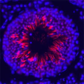
ESGP in Mouse Testis. ESGP was detected in perfusion fixed frozen sections of mouse testis using Sheep Anti-Mouse ESGP Antigen Affinity-purified Polyclonal Antibody (Catalog # AF4580) at 1.7 µg/mL overnight at 4 °C. Tissue was stained using the Northern-Lights™ 557-conjugated Anti-Sheep IgG Secondary Antibody (red; Catalog # NL010) and counterstained with DAPI (blue). Specific staining was localized to late spermatids. View our protocol for Fluorescent IHC Staining of Frozen Tissue Sections.
 View Larger
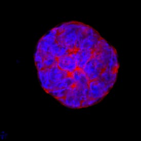
ESGP in D3 Mouse Cell Line. ESGP was detected in immersion fixed D3 mouse embryonic stem cell line using Sheep Anti-Mouse ESGP Antigen Affinity-purified Polyclonal Antibody (Catalog # AF4580) at 10 µg/mL for 3 hours at room temperature. Cells were stained using the NorthernLights™ 557-conjugated Anti-Sheep IgG Secondary Antibody (red; Catalog # NL010) and counterstained with DAPI (blue). Specific staining was localized to cytoplasm and cell secretion. View our protocol for Fluorescent ICC Staining of Stem Cells on Coverslips.
Preparation and Storage
- 12 months from date of receipt, -20 to -70 °C as supplied.
- 1 month, 2 to 8 °C under sterile conditions after reconstitution.
- 6 months, -20 to -70 °C under sterile conditions after reconstitution.
Background: ESGP
ESGP (ES cell and germ cell-specific protein) is a presumably secreted polypeptide that belongs to no known protein family. It is expressed specifically in pluripotential cell types, and does not appear to be involved in differentiation. Mouse ESGP precursor is 84 amino acids (aa) in length. It contains a 25 aa signal sequence with a 59 aa mature segment. There are no N-linked glycosylation sites. One potential 108 aa variant shows an alternate start site 24 aa upstream of the standard start site.
Product Datasheets
Citations for Mouse ESGP Antibody
R&D Systems personnel manually curate a database that contains references using R&D Systems products. The data collected includes not only links to publications in PubMed, but also provides information about sample types, species, and experimental conditions.
13
Citations: Showing 1 - 10
Filter your results:
Filter by:
-
Myomaker and Myomerger Work Independently to Control Distinct Steps of Membrane Remodeling during Myoblast Fusion
Authors: Evgenia Leikina, Dilani G. Gamage, Vikram Prasad, Joanna Goykhberg, Michael Crowe, Jiajie Diao et al.
Developmental Cell
-
Filopodia powered by class x myosin promote fusion of mammalian myoblasts
Authors: Hammers DW, Hart CC, Matheny MK et al.
eLife
-
Survival motor neuron deficiency slows myoblast fusion through reduced myomaker and myomixer expression
Authors: Nikki M. McCormack, Eric Villalón, Coralie Viollet, Anthony R. Soltis, Clifton L. Dalgard, Christian L. Lorson et al.
Journal of Cachexia, Sarcopenia and Muscle
-
Formation of Aberrant Myotubes by Myoblasts Lacking Myosin VI Is Associated with Alterations in the Cytoskeleton Organization, Myoblast Adhesion and Fusion
Authors: Lilya Lehka, Małgorzata Topolewska, Dominika Wojton, Olena Karatsai, Paloma Alvarez-Suarez, Paweł Pomorski et al.
Cells
-
Interleukin‐4 administration improves muscle function, adult myogenesis, and lifespan of colon carcinoma‐bearing mice
Authors: Domiziana Costamagna, Robin Duelen, Fabio Penna, Detlef Neumann, Paola Costelli, Maurilio Sampaolesi
Journal of Cachexia, Sarcopenia and Muscle
-
IL-4 Signaling Promotes Myoblast Differentiation and Fusion by Enhancing the Expression of MyoD, Myogenin, and Myomerger
Authors: Kurosaka, M;Hung, YL;Machida, S;Kohda, K;
Cells
Species: Mouse
Sample Types: Cell Lysates
Applications: Western Blot -
Phosphatidylserine orchestrates Myomerger membrane insertions to drive myoblast fusion
Authors: DG Gamage, K Melikov, P Munoz-Tell, TJ Wherley, LC Focke, E Leikina, E Huffman, J Diao, DJ Kojetin, V Prasad, LV Chernomord, DP Millay
Proceedings of the National Academy of Sciences of the United States of America, 2022-09-12;119(38):e2202490119.
Species: Mouse
Sample Types: Whole Cells
Applications: ICC -
LncRNA OIP5-AS1-directed miR-7 degradation promotes MYMX production during human myogenesis
Authors: JH Yang, MW Chang, D Tsitsipati, X Yang, JL Martindale, R Munk, A Cheng, E Izydore, PR Pandey, Y Piao, K Mazan-Mamc, S De, K Abdelmohse, M Gorospe
Nucleic Acids Research, 2022-07-08;0(0):.
Species: Human
Sample Types: Cell Lysates
Applications: Western Blot -
Magnesium Homeostasis in Myogenic Differentiation-A Focus on the Regulation of TRPM7, MagT1 and SLC41A1 Transporters
Authors: M Zocchi, L Locatelli, GV Zuccotti, A Mazur, D Béchet, JA Maier, S Castiglion
International Journal of Molecular Sciences, 2022-01-31;23(3):.
Species: Mouse
Sample Types: Cell Lysates
Applications: Western Blot -
Filopodia powered by class x myosin promote fusion of mammalian myoblasts
Authors: Hammers DW, Hart CC, Matheny MK et al.
eLife
-
Fusogenic micropeptide Myomixer is essential for satellite cell fusion and muscle regeneration
Authors: P Bi, JR McAnally, JM Shelton, E Sánchez-Or, R Bassel-Dub, EN Olson
Proc. Natl. Acad. Sci. U.S.A., 2018-03-26;0(0):.
Species: Mouse
Sample Types: Tissue Homogenates
Applications: Western Blot -
The microprotein Minion controls cell fusion and muscle formation
Authors: Q Zhang, AA Vashisht, J O'Rourke, SY Corbel, R Moran, A Romero, L Miraglia, J Zhang, E Durrant, C Schmedt, SC Sampath, SC Sampath
Nat Commun, 2017-06-01;8(0):15664.
Species: Mouse
Sample Types: Whole Cells, Whole Tissue
Applications: ICC, IHC -
Myomerger induces fusion of non-fusogenic cells and is required for skeletal muscle development
Authors: ME Quinn, Q Goh, M Kurosaka, DG Gamage, MJ Petrany, V Prasad, DP Millay
Nat Commun, 2017-06-01;8(0):15665.
Species: Mouse
Sample Types: Tissue Homogenates
Applications: Western Blot
FAQs
No product specific FAQs exist for this product, however you may
View all Antibody FAQsIsotype Controls
Reconstitution Buffers
Secondary Antibodies
Reviews for Mouse ESGP Antibody
There are currently no reviews for this product. Be the first to review Mouse ESGP Antibody and earn rewards!
Have you used Mouse ESGP Antibody?
Submit a review and receive an Amazon gift card.
$25/€18/£15/$25CAN/¥75 Yuan/¥2500 Yen for a review with an image
$10/€7/£6/$10 CAD/¥70 Yuan/¥1110 Yen for a review without an image


