Mouse Ki67/MKI67 Antibody Summary
Applications
Please Note: Optimal dilutions should be determined by each laboratory for each application. General Protocols are available in the Technical Information section on our website.
Scientific Data
 View Larger
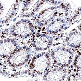
Detection of Ki67/MKI67 in Mouse Colon. Ki67/MKI67 was detected in immersion fixed paraffin-embedded sections of mouse colon using Mouse Anti-Mouse Ki67/MKI67 Monoclonal Antibody (Catalog # MAB11612) at 5 µg/ml overnight at 4 °C. Before incubation with the primary antibody, tissue was subjected to heat-induced epitope retrieval using VisUCyte Antigen Retrieval Reagent-Basic (Catalog # VCTS021). Tissue was stained using the HRP-conjugated Anti-Rat IgG Secondary Antibody (Catalog # HAF005) and counterstained with hematoxylin (blue). Specific staining was localized to the nucleus. View our protocol for Chromogenic IHC Staining of Paraffin-embedded Tissue Sections.
 View Larger
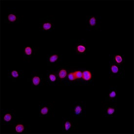
Detection of Ki67/MKI67 in Raw264.7. Ki67/MKI67 was detected in immersion fixed RAW264.7 cells using Mouse Anti-Mouse Ki67/MKI67 Monoclonal Antibody (Catalog # MAB11612) at 8 µg/ml for 3 hours at room temperature. Cells were stained using the NorthernLights™ 557-conjugated Anti-Rat IgG Secondary Antibody (red; Catalog # NL013) and counterstained with DAPI (blue). Specific staining was localized to the nucleus. View our protocol for Fluorescent ICC Staining of Cells on Coverslips.
Reconstitution Calculator
Preparation and Storage
- 12 months from date of receipt, -20 to -70 °C as supplied.
- 1 month, 2 to 8 °C under sterile conditions after reconstitution.
- 6 months, -20 to -70 °C under sterile conditions after reconstitution.
Background: Ki67/MKI67
MKI67 (also Ki67 and TSG126) is a 350-370 kDa nuclear protein that belongs to a molecular group comprised of mitotic chromosome-associated proteins. Ki67 was originally recognized as an antigen associated with the monoclonal Ki67 antibody raised against Hodgkin's lymphoma nuclear material. Ki67 is contextually expressed, being potentially found in all cells that are not in the Go phase of the cell cycle. Thus, MKI67 qualifies as a cell proliferation marker. Functionally, Ki67 is known to interact with 160 kDa Hklp2, a protein that promotes centrosome separation and spindle bipolarity. It also directly interacts with NIFK, and apparently binds to UBF, thus playing a role in rRNA synthesis. Mouse MKI67 is 3177 amino acids (aa) in length. It contains one FHA domain (aa 8-101), followed by sixteen 120 aa repeats (aa 993-2872). There are two potential isoform variants. One isoform shows a 19 aa substitution for 1120-3177, while a second isoform contains a deletion of aa 1169-1409. Over aa 3053‑3177, mouse Ki67 shares 46% and 74% aa sequence identity with the human and rat orthologs to Ki67, respectively.
Product Datasheets
FAQs
No product specific FAQs exist for this product, however you may
View all Antibody FAQsReviews for Mouse Ki67/MKI67 Antibody
There are currently no reviews for this product. Be the first to review Mouse Ki67/MKI67 Antibody and earn rewards!
Have you used Mouse Ki67/MKI67 Antibody?
Submit a review and receive an Amazon gift card.
$25/€18/£15/$25CAN/¥75 Yuan/¥2500 Yen for a review with an image
$10/€7/£6/$10 CAD/¥70 Yuan/¥1110 Yen for a review without an image

