 |
| View Graphic Protocol |
The following immunohistochemistry (IHC) protocol has been developed and optimized by R&D Systems’ immunohistochemistry (IHC)/immunocytochemistry (ICC) laboratory for immunofluorescence IHC experiments using frozen tissues samples
This cryosection immunofluorescence protocol provides a basic guide for the fixation, cryostat sectioning, and staining of frozen tissue samples. Each investigator must determine the precise experimental conditions required to generate a strong and specific signal for each antigen of interest. If R&D Systems' primary antibodies are employed, please refer to the product data sheets to obtain the recommended working dilutions. In the frozen section staining protocol, signal visualization is achieved using R&D Systems' fluorescent secondary antibodies. For all other detection reagents, please follow the manufacturer's instructions.
Please read IHC protocol in its entirety before beginning.
Immunohistochemistry Protocol for Fixing and Sectioning Frozen Tissues
Reagents Required
- O.C.T. Embedding Compound
- Formaldehyde Fixative Solution: 85 mM Na2HPO4, 75 mM KH2P04, 4% paraformaldehyde, and 14% (v/v) saturated picric acid, pH 6.9. (Picric acid is optional).
- Sucrose Solution: 130 mM Na2HPO4, 30 mM KH2P04, 10% (w/v) sucrose, 0.01% sodium azide and 0.03% Bacitracin, pH 7.2
- Dry Ice
- Isopentane
IHC Cryosection Protocol for Fixing and Sectioning of Frozen Tissues
This technique utilizes formaldehyde-based fixation before the tissue is frozen and sectioned using a cryostat.
- To preserve tissue morphology and retain the antigenicity of the target molecules, fix the tissue by vascular perfusion with 500 - 700 mL of Formaldehyde Fixative Solution.
- Perfuse the animal with 400 mL of Sucrose Solution.
Note: When it is not possible to fix by perfusion, dissected tissue may be fixed by immersion in a 10% formalin solution for 4 to 8 hours at room temperature. It is commonly accepted that the volume of fixative should be 50 times greater than the size of the immersed tissue. Avoid fixing the tissue for greater than 24 hours since tissue antigens may either be masked or destroyed.
Note: R&D Systems scientists perfuse fix all rodent tissue with the exception of lung, spleen, and embryonic tissue, which are immersion fixed. - Dissect the tissue, mount in OCT embedding compound, and freeze at -20 to -80 °C.
- Cut 5-15 µm thick tissue sections using a cryostat.
Note: The suggested cryostat temperature is between -15 and -23 °C. The section will curl if the specimen is too cold. If it is too warm, it will stick to the knife. - Thaw-mount the sections onto gelatin-coated histological slides. Slides are pre-coated with gelatin to enhance adhesion of the tissue. Please refer to the Protocol for the Preparation of Gelatin-coated Slides for Histological Tissue Sections for instructions on how to prepare gelatin-coated slides.
- Dry the slides for 30 minutes on a slide warmer at 37 °C. Slides with mounted frozen tissue sections can be stored at -20 to -70 °C for up to 12 months.
Immunohistochemistry Protocol for Cryopreservation of Tissues Prior to Fixation
This method utilizes frozen tissues that are fixed after snap-freezing and sectioning with a cryostat.
- Immediately snap freeze fresh tissue in isopentane mixed with dry ice, and keep at -70 °C. Do not allow frozen tissue to thaw before cutting.
- Embed the tissue completely in OCT compound prior to cryostat sectioning.
- Cut frozen sections at 5-10 µm and mount on gelatin-coated histological slides.
Note: The suggested cryostat temperature is between -15 and -23 °C. The section will curl if the specimen is too cold. If it is too warm, it will stick to the knife. - Air dry the frozen sections for 30 minutes at room temperature to prevent sections from falling off the slides during antibody incubations.
Note: Slides can be stored unfixed for several months at -70 °C. Frozen tissue samples saved for later analysis should be stored intact. - Immediately add 50 µL of ice-cold Fixation Buffer to each tissue section upon removal from the freezer.
- Fix for 8 minutes at 2-8 °C or, optimally, at -20 °C for 20 minutes.
Immunohistochemistry Protocol for Immunofluorescence Cryosection Staining
Reagents Required
- Primary Antibodies
- NorthernLightsTM Secondary Antibodies (or equivalent)
- Wash Buffer: 1X PBS (0.145 M NaCl, 0.0027 M KCl, 0.0081 M Na2HPO4, 0.0015 M KH2PO4, pH 7.4)
- Incubation Buffer: 1% bovine serum albumin, 1% normal donkey serum, 0.3% Triton® X-100, and 0.01% sodium azide in PBS
- Blocking buffer: 1% horse serum in PBS
- DAPI (4',6-diamidino-2-phenylindole) solution: Add 1 µL of 14.3 mM stock for every 5 mL of PBS. Store any unused DAPI at 2-8 °C, wrapped in aluminum foil
- Anti-Fade Mounting Medium
- Antigen Retrieval Reagents (if required; Protocol for Heat-induced Epitope Retrieval (HEIR))
Immunohistochemistry (IHC) Protocol for Frozen Tissue Sections
- When staining cryostat sections stored in a freezer, thaw the slides at room temperature for 10-20 minutes.
- Rehydrate the slides in wash buffer for 10 minutes. Drain the excess wash buffer.
Note: Excessive fixation may result in the masking of an epitope and strong non-specific background signal that can obscure specific labeling. If necessary, an antigen retrieval protocol can be performed at this time. However, many antigen retrieval techniques are too harsh for cryostat-cut tissue sections. - Surround the tissue with a hydrophobic barrier using a barrier pen.
- Block non-specific staining between the primary antibodies and the tissue, by incubating in blocking buffer (1% horse serum in PBS) for 30 minutes at room temperature.
- Apply primary antibodies diluted in Incubation Buffer according to the manufacturer’s instructions. For immunofluorescence staining of frozen tissue sections using R&D Systems antibodies, it is recommended to incubate overnight at 2-8 °C. This incubation regime allows for optimal specific binding of antibodies to tissue targets and reduces non-specific background staining. These variables may need to be optimized for your system.
Note: Appropriate controls are critical for the accurate interpretation of IHC/ICC results. All IHC/ICC experiments should include a negative control using the incubation buffer with no primary antibody to identify non-specific staining of the secondary reagents. Additional controls can be employed to support the specificity of staining generated by the primary antibody. These include absorption controls, isotype controls (for monoclonal primary antibodies), and tissue type controls. - Wash slides 3 times for fifteen minutes each in wash buffer.
- Incubate with the NorthernLights™ secondary antibody diluted in Incubation Buffer according to the manufacturer’s instructions. Recommended incubation with NorthernLights secondary antibody is for 30-60 minutes at room temperature. From this step forward samples should be protected from light.
Note: R&D Systems NorthernLights™ fluorescent secondary antibodies and streptavidin conjugates are bright, resistant to photobleaching and are ideal for multi-color fluorescence microscopy.
Note: If a biotinylated antibody was used in step 5, apply the appropriate NorthernLights™ Streptavidin conjugate in step 7. - Wash slides 3 times for fifteen minutes each in Wash Buffer.
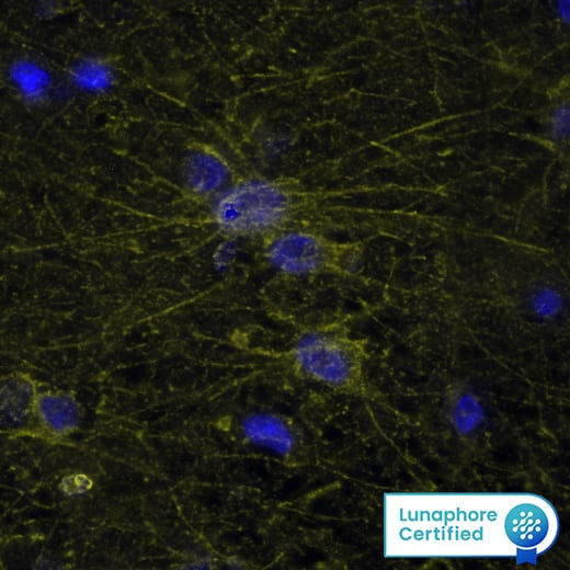
- Add 300 µL of the diluted DAPI solution to each well and incubate 2-5 minutes at room temperature. DAPI binds to DNA and is a convenient nuclear counterstain. It has an absorption maximum at 358 nm and fluoresces blue at an emission maximum of 461 nm.
Note: DAPI counterstain can obscure visualization of targets localized in cell nuclei. - Rinse 1 time with PBS.
- Mount with an anti-fade mounting media.
- Visualize using a fluorescence microscope.
Note: Initial IHC/ICC studies often require further optimization and/or additional troubleshooting steps.
Triton is a registered trademark of Dow Chemical.
Related IHC/ICC Resources
- Immunohistochemistry (IHC) Handbook
- Troubleshooting Guide for Immunohistochemistry and Immunocytochemsitry
- Protocol for Making a 4% Formaldehyde Solution in PBS
- IHC/ICC Protocol Guide
- Designing a Successful IHC/ICC Experiment
- All IHC/ICC protocols
- Preventing Non-specific Staining Protocol


