Tumors recruit immunosuppressive immune cells which create a microenvironment that inhibits anti-tumor responses and enables tumor growth. Monitoring the spatial and temporal dynamics of tumor infiltrating lymphocytes (TILs) and other immune cells in the tumor microenvironment is crucial for the development of more effective immune checkpoint blockade strategies. R&D Systems offers a wide selection of flow cytometry antibodies and kits for phenotyping immune cells and cell selection kits to isolate them for downstream studies. Immune cell phenotyping is an important step in the cell therapy workflow. To view our complete solutions for Cell and Gene Therapy manufacturing, including all GMP-grade reagents and analytical instrumentation, visit bio-techne.com.
Immune Checkpoint Blockade Homepage
Flow Cytometry Antibodies for Immune Cell Markers
Click the buttons below to view select markers commonly used to identify each cell type. The lists of markers are general and it is recognized that there are different subtypes of many of the cell types below (e.g. macrophages and NKT cells) with opposing functions in the anti-tumor immune response. Molecules are linked directly to flow cytometry antibodies making it easy to find what you need.
| B Cell Markers | T Cell Markers | Th17 Cell Markers | Regulatory T Cell Markers | NK Cell Markers |
| NKT Cell Markers | Monocyte Markers | Macrophage Markers | Dendritic Cell Markers | MDSC Markers |
Multi-color Flow Cytometry Kits
Identify cell subsets with our rapid and efficient multi-color flow cytometry kits that simultaneously stain multiple targets in a single step. Our kits include fluorochrome-conjugated antibodies, isotype controls for each antibody, and all necessary buffers.
| Regulatory T Cell Kits | Th17 Cell Kits | Dendritic Cell Kits | View All Flow Cytometry Kits |
 |
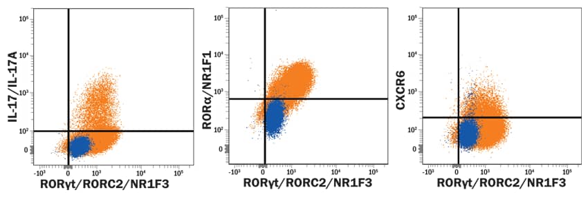
| Th17 cells were harvested and stained with the indicated antibodies following the procedure described in the product description (Catalog # FMC007B). Live, single, CD4+ cells are shown in the dot plots (determined using a fixable viability dye, doublet exclusion, and staining with anti-human CD4-Alexa Fluor® 594). Dot plots show relative RORγt+, IL-17A+, RORα+, and CXCR6+ cells in CD4+ resting (blue dots, lower left) and Th17-differentiated (orange dots, upper right quadrants). Quadrant markers were set based on isotype controls. |
MagCellect™ Cell Selection Kits
Our cell selection kits utilize MagCellect technology based on the use of ferrofluids or magnetic nanoparticles that have no magnetic memory (i.e. superparamagnetic). This feature protects purified cell populations against magnetic forces which may affect cell viability and interfere with downstream applications.
| CD3+ T Cells | CD4+ T Cells | CD8+ T Cells | Regulatory T Cells |
| NK Cells | CD14+ Monocytes/Macrophages | B Cells | View All Cell Selection Kits |
Immune Cell Markers
| B Cell Markers | |
| Human | Mouse |
| CD19 | CD19 |
| MS4A1/CD20 | |
| T Cell Markers | |
| Human | Mouse |
| CD3 | CD3 |
| CD4 | CD4 |
| CD8 | CD8 |
| Th17 Cell Markers | |
| Human | Mouse |
| Regulatory T Cell Markers | |
| Human | Mouse |
| NK Cell Markers | |
| Human | Mouse |
| NKT Cell Markers | |
| Human | Mouse |
|
V alpha 24 |
CD1d tetramer |
|
V beta 11 |
TCR beta |
|
CD1d tetramer |
|
|
V alpha 14 |
|
| Monocyte Markers | |
| Human | Mouse |
|
Ly-6C |
|
| Macrophage Markers | |
| Human | Mouse |
|
CD68/SR-D1 |
|
| Dendritic Cell Markers | |
| Human | Mouse |
| MDSC Markers | |
| Human | Mouse |
|
Ly-6C |
|