Activated T cells in the tumor microenvironment can induce the expression of B7-H1/PD-L1 on cancer cells. B7-H1/PD-L1 then negatively regulates effector T cell responses through its interaction with PD-1 on activated T cells. Importantly, B7-H1/PD-L1 expression on cancer cells has been positively correlated with responsiveness to B7-H1/PD-L1 blockade. Trust the antibodies and Luminex® Screening Assays offered by R&D Systems to monitor the expression of B7-H1/PD-L1 and other checkpoint molecules in your research.
Immune Checkpoint Blockade Homepage
Detect B7-H1/PD-L1 Expression
R&D Systems offers high-quality antibodies for the detection of B7-H1/PD-L1 by immunohistochemistry (IHC), flow cytometry, and Western blot. Our antibodies are manufactured and carefully tested with quality and performance as the top priorities. Learn more about what makes our antibodies the best.
| B7-H1/PD-L1 IHC Antibodies | B7-H1/PD-L1 Flow Cytometry Antibodies | B7-H1/PD-L1 Western Blot Antibodies |
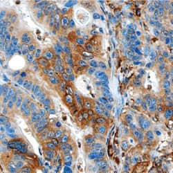
 Detection of B7-H1/PD-L1 in Human Colon Cancer Tissue. B7-H1/PD-L1 was detected in formalin fixed paraffin-embedded sections of human colon cancer using Mouse Anti-Human B7-H1/PD-L1 Monoclonal Antibody (Catalog # MAB1561). Tissue was stained using the Anti-Mouse HRP-DAB Cell & Tissue Staining Kit (brown; Catalog # CTS002) and counterstained with hematoxylin (blue). Specific staining was observed in the cytoplasm. View our protocol for Chromogenic IHC Staining of Paraffin-embedded Tissue Sections. |
 Detection of B7-H1/PD-L1 on Mouse Splenocytes. Mouse splenocytes were stained with Goat Anti-Mouse B7-H1/PD-L1 Fluorescein-conjugated Antigen Affinity-purified Polyclonal Antibody (Catalog # FAB1019F, filled histogram) or isotype control antibody (Catalog # IC108F, open histogram). View our protocol for Staining Membrane-associated Proteins. |
 Detection of B7-H1/PD-L1 by Western blot. Lysates of human placenta tissue were separated by SDS-PAGE. The PVDF membrane was probed with Goat Anti-Human B7-H1/PD-L1 Antigen Affinity-purified Polyclonal Antibody (Catalog # AF156) followed by HRP-conjugated Anti-Goat IgG Secondary Antibody (Catalog # HAF017). This experiment was conducted under reducing conditions and using Immunoblot Buffer Group 1. |
View IHC Antibodies for More T Cell Co-inhibitory Molecules
Get More Data From Your Sample
Do you need to determine the expression levels of more than one analyte? Utilize our Luminex® Screening Assays to simultaneously analyze the expression levels of up to 99 other human analytes in combination with B7-H1/PD-L1. Our Screening Assays are cost-effective, accurate, flexible, and require a small sample volume (‹50 µL).
| Luminex Assays |  |
Luminex Assay Customization Tool |