Mouse IgG1 Alexa Fluor® 488-conjugated Antibody Summary
Applications
Please Note: Optimal dilutions should be determined by each laboratory for each application. General Protocols are available in the Technical Information section on our website.
Scientific Data
 View Larger
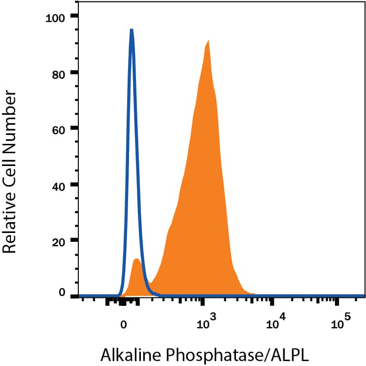
Detection of Mouse IgG Control by Flow Cytometry BG01V human embryonic stem cells were stained with Mouse Anti-Human Alkaline Phosphatase/ALPL Alexa Fluor® 488-conjugated Monoclonal Antibody (Catalog # FAB1448G, filled histogram) or Mouse IgG Alexa Fluor® 488-conjugated Isotype Control Antibody (Catalog # IC002G, open histogram). View our protocol for Staining Membrane-associated Proteins.
Reconstitution Calculator
Preparation and Storage
- 12 months from date of receipt, 2 to 8 °C as supplied.
Background: IgG1
R&D Systems offers a range of secondary antibodies and controls for flow cytometry, immunohistochemistry, and Western blotting. We provide species-specific secondary antibodies that are available with a variety of conjugated labels.
Our NorthernLights fluorescent secondary antibodies are bright and resistant to photobleaching. We are currently offering secondary antibodies recognizing mouse, rat, goat, sheep, and rabbit IgG as well as chicken IgY. These reagents are available with three distinct excitation and emission maxima, making them ideal for multi-color fluorescence microscopy.
Product Datasheets
Citations for Mouse IgG1 Alexa Fluor® 488-conjugated Antibody
R&D Systems personnel manually curate a database that contains references using R&D Systems products. The data collected includes not only links to publications in PubMed, but also provides information about sample types, species, and experimental conditions.
13
Citations: Showing 1 - 10
Filter your results:
Filter by:
-
Characterization of the Filovirus-Resistant Cell Line SH-SY5Y Reveals Redundant Role of Cell Surface Entry Factors
Authors: FJ Zapatero-B, E Dietzel, O Dolnik, K Döhner, R Costa, B Hertel, B Veselkova, J Kirui, A Klintworth, MP Manns, S Pöhlmann, T Pietschman, T Krey, S Ciesek, G Gerold, B Sodeik, S Becker, T von Hahn
Viruses, 2019-03-19;11(3):.
-
A Multi-Color Flow Cytometric Assay for Quantifying Dinutuximab Binding to Neuroblastoma Cells in Tumor, Bone Marrow, and Blood
Authors: Keyel, ME;Furr, KL;Kang, MH;Reynolds, CP;
Journal of clinical medicine
Species: Human
Sample Types: Whole Cells
Applications: Flow Cytometry -
Senescent mesenchymal stem/stromal cells in pre-metastatic bone marrow of untreated advanced breast cancer patients
Authors: Francisco Raúl RA贚 BORZONE, María Belén BEL蒒 GIORELLO, Leandro Marcelo MARCELO MARTINEZ, María Cecilia CECILIA SANMARTIN, LEONARDO FELDMAN, FEDERICO DIMASE et al.
Oncology Research
-
A CRISPRi/a platform in human iPSC-derived microglia uncovers regulators of disease states
Authors: NM Dräger, SM Sattler, CT Huang, OM Teter, K Leng, SH Hashemi, J Hong, G Aviles, CD Clelland, L Zhan, JC Udeochu, L Kodama, AB Singleton, MA Nalls, J Ichida, ME Ward, F Faghri, L Gan, M Kampmann
Nature Neuroscience, 2022-08-11;0(0):.
Species: Human
Sample Types: Whole Cells
Applications: Flow Cytometry -
The Quest for Anti-?-Synuclein Antibody Specificity-Lessons Learnt From Flow Cytometry Analysis
Authors: Leupold L, Sigutova V, Gerasimova E et al.
Frontiers in neurology
-
Differential Marker Expression between Keratinocyte Stem Cells and Their Progeny Generated from a Single Colony
Authors: D Ali, D Alhattab, H Jafar, M Alzubide, N Sharar, S Bdour, A Awidi
International Journal of Molecular Sciences, 2021-10-06;22(19):.
Species: Human
Sample Types: Whole Cells
Applications: Flow Cytometry -
High-throughput single-EV liquid biopsy: Rapid, simultaneous, and multiplexed detection of nucleic acids, proteins, and their combinations
Authors: J Zhou, Z Wu, J Hu, D Yang, X Chen, Q Wang, J Liu, M Dou, W Peng, Y Wu, W Wang, C Xie, M Wang, Y Song, H Zeng, C Bai
Sci Adv, 2020-11-20;6(47):.
Species: Human
Sample Types: Extracellular Vesicles
Applications: ICC -
Human NK Cells Lyse Th2-Polarizing Dendritic Cells via NKp30 and DNAM-1
Authors: K Walwyn-Bro, K Guldevall, M Saeed, D Pende, B Önfelt, AS MacDonald, DM Davis
J. Immunol., 2018-08-17;0(0):.
Species: Human
Sample Types: Whole Cells
Applications: Flow Cytometry -
Dual role of DR5 in death and survival signaling leads to TRAIL resistance in cancer cells
Authors: Y Shlyakhtin, V Pavet, H Gronemeyer
Cell Death Dis, 2017-08-31;8(8):e3025.
Species: Human
Sample Types: Whole Cells
Applications: Flow Cytometry -
Induction and Differentiation of IL-10-Producing Regulatory B Cells from Healthy Blood Donors and Rheumatoid Arthritis Patients.
Authors: Banko Z, Pozsgay J, Szili D, Toth M, Gati T, Nagy G, Rojkovich B, Sarmay G
J Immunol, 2017-01-13;198(4):1512-1520.
Species: Human
Sample Types: Whole Cells
Applications: Flow Cytometry Control -
Induction of indoleamine 2,3-dioxygenase by Borrelia burgdorferi in human immune cells correlates with pathogenic potential.
Authors: Love A, Schwartz I, Petzke M
J Leukoc Biol, 2014-11-24;97(2):379-90.
Species: Human
Sample Types: Whole Cells
Applications: Flow Cytometry -
Intrapatient variations in type 1 diabetes-specific iPS cell differentiation into insulin-producing cells.
Authors: Thatava T, Kudva Y, Edukulla R, Squillace K, De Lamo J, Khan Y, Sakuma T, Ohmine S, Terzic A, Ikeda Y
Mol Ther, 2012-11-27;21(1):228-39.
Species: Human
Sample Types: Whole Cells
Applications: Flow Cytometry Control -
Cellular plasticity and immune microenvironment of malignant pleural effusion are associated with EGFR-TKI resistance in non-small-cell lung carcinoma
Authors: Jeong H, Lee H, Kim H et al.
iScience
FAQs
No product specific FAQs exist for this product, however you may
View all Isotype Control FAQsReviews for Mouse IgG1 Alexa Fluor® 488-conjugated Antibody
Average Rating: 4.8 (Based on 4 Reviews)
Have you used Mouse IgG1 Alexa Fluor® 488-conjugated Antibody?
Submit a review and receive an Amazon gift card.
$25/€18/£15/$25CAN/¥75 Yuan/¥2500 Yen for a review with an image
$10/€7/£6/$10 CAD/¥70 Yuan/¥1110 Yen for a review without an image
Filter by:
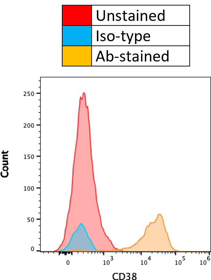
Use as Iso-type Ctrl
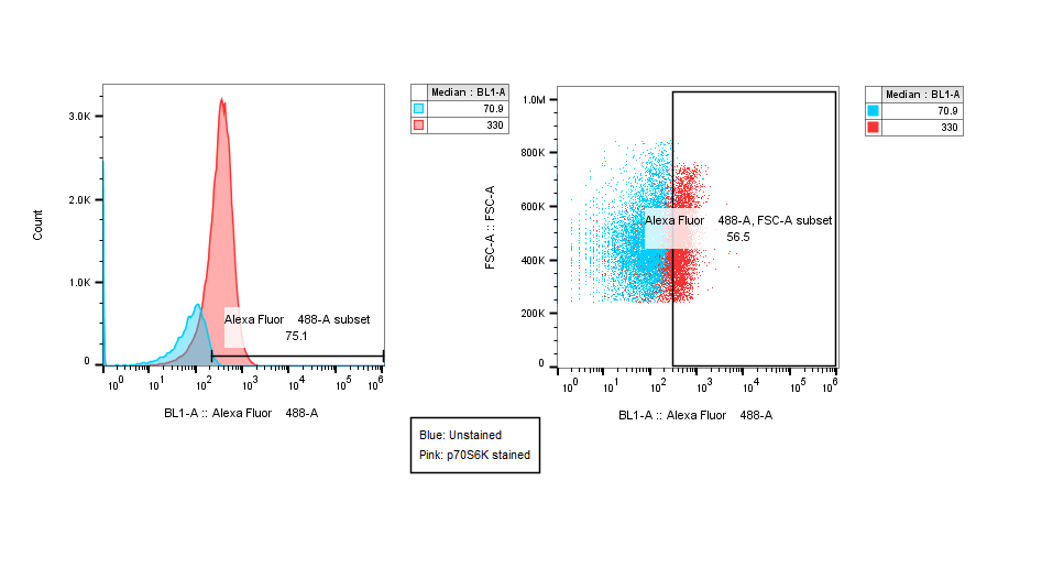
MCF, cells were fixed and permeabilized and the stained with the antibody and the intracellular expression was measured by Flow Cytometry
B16F10 cells were stained extracellularly with the antibodies and Expression was measured by Flow cytometry.


