Conjugated Primary Antibodies

Conjugated Antibodies

Search Conjugated Antibodies

Conjugated Primary Antibodies

R&D Systems offers a wide variety of conjugated primary antibodies allowing you to analyze multiple targets with ease. Our extensive selection of conjugated antibodies in a variety of fluorescent and enzymatic labels allows you to customize your experiment. We offer bright conjugates for low abundance analytes and subdued dyes for high abundance targets. Find your antibody panel today!

See All Primary Conjugated Antibodies


What Are Conjugated Antibodies?

A conjugated antibody is a polyclonal or monoclonal antibody that has a molecule attached which can be used to create a detectable signal. Detection can be visualized by color-generation, fluorescence, or other signals. Conjugated antibodies are used in a wide range of applications including flow cytometry, immunohistochemistry, immunocytochemistry, western blot, and ELISA.

Types of Detection Methods

There are two types of detection methods used with antibodies, direct and indirect.

Direct detection uses a primary antibody that is directly conjugated to a label. By conjugating primary antibodies, you can directly detect the target without use of secondary antibodies. The advantages of direct detection include ease of use for multicolor staining and eliminating the concerns regarding non-specific binding of the secondary antibody. It is ideal to use directly conjugated antibodies when the target of interest is in high abundance, or the conjugate signal can compensate for low target expression levels.

Indirect detection uses a primary antibody that is unconjugated and a conjugated secondary antibody that is specific for the host species of the primary antibody. Indirect detection is the preferred method if the target of interest has a lower level of expression. By using secondary antibodies, this allows signal amplification due to the secondary antibodies carrying multiple labels that can bind to the primary antibody. In general, indirect detection methods generally have a higher level of sensitivity and generate a more intense signal.

direct and indirect detection for antibodies


Fluorescent Dyes

Fluorescence detection is the emission of light by a molecule that has absorbed light at a shorter wavelength. Fluorescent dyes use fluorochromes that have an absorption and emission wavelength. They emit a photon at one wavelength and when excited by the presence of light, excite at another. By using different fluorescently conjugated antibodies, you can detect multiple targets at once. It is important to take note of the excitation and emission spectra if using multiple fluorescent dyes. You will want to avoid overlapping emission spectrums when performing co-staining. Select a brighter dye, with a higher extinction coefficient, for lower abundance targets. Fluorescent conjugated antibodies can be used in many different techniques including flow cytometry, FACS, and immunofluorescence. R&D Systems offers a wide variety of antibodies that are directly conjugated to a fluorescent molecule.


Fluorescent conjugated antibodies

Cytokeratin 18 was detected in formaldehyde fixed HeLa human cervical epithelial carcinoma cell line using Mouse Anti-Human Cytokeratin 18 Alexa Fluor® 488 conjugated Monoclonal Antibody (Catalog # IC7619G).

Fluorescent conjugated antibodies

SSEA 1 was detected in immersion fixed D3 mouse embryonic stem cell line using Mouse Anti-Human/Mouse SSEA 1 Alexa Fluor® 594 conjugated Monoclonal Antibody (Catalog # IC2155T).

Fluorescent conjugated antibodies

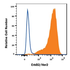
MDA-MB-453 human breast cancer cell line was stained with Mouse Anti-Human ErbB2/Her2 PE conjugated Monoclonal Antibody (Catalog # FAB1129P).


 Available Fluorescent Conjugation Options by Laser 

UV 360nm

Violet 405nm

Blue 488nm

Yellow-Green 561nm

Red 633nm

Additional conjugation options are available through Novus Biologicals


Chromogenic and Biotin Labels

Antibodies can also be conjugated to a label that provides chromogenic detection instead of fluorescence. When an antibody is conjugated to an enzyme such as horseradish peroxidase (HRP) or alkaline phosphatase, the enzyme reacts to its substrate and an insoluble colored product is localized to the sites of antigen expression. Chromogenic labeled antibodies are great options for ELISA , Western blot, and Immunohistochemistry.


Biotin conjugated antibodies are recommended to be used when target expression is low. When a biotinylated antibody is used, the signal can be significantly amplified by subsequent incubation with a labeled streptavidin-biotin (LSAB Method). This due to the ability of streptavidin to bind up to four biotins per molecule. Fluorescent or chromogenic conjugated streptavidin is used to detect the biotin on the primary antibody and produce a signal.


An alternative option to the biotin-streptavidin amplification complex, is R&D Systems’ VisUCyte HRP Polymer. The VisUCyte HRP Polymer is a biotin-free detection reagent that eliminates the need to perform additional quenching of endogenous biotin and avidin present in some tissues. With fewer blocking and incubation steps, researchers can achieve specific staining in cells and tissues faster compared to the traditional streptavidin-biotin-HRP procedure.

VisUCyte HRP Polymer


Isotype Controls and Secondary Reagents

While we strive to offer the entire menu of conjugates for our antibodies, it is not always feasible. Some antibodies do not conjugate well to certain labels. The use of a secondary reagent can overcome these difficulties. We offer secondary antibodies conjugated to fluorescent and chromogenic labels.

Secondary Antibodies


To confirm staining, it is recommended to use an appropriate control. Isotype controls can be used to determine if non-specific staining is present. Cells can have natural fluorescence and non-specific binding can occur, so it is important to confirm staining that is observed. Isotype control antibodies lack specificity to a target of interest but have the same host species, class, and conjugate as the primary antibody.

Isotype Controls