Introduction to Secondary Antibodies

Secondary Antibodies

Search Secondary Antibodies

Secondary antibodies are a powerful tool that can help amplify signal and increase sensitivity in an assay. Since multiple secondary antibodies are able to detect a primary antibody, signal is increased and in turn creates a more sensitive assay. Secondary antibodies can also provide flexibility in multiple labeling experiments by having the ability to pick from a variety of diverse conjugation options to fit the specific needs of the assay. 

R&D Systems offers over 90 different secondary antibody options specific for a variety of species and conjugated to fluorescent and chromogenic labels. Our secondary antibodies have been validated in-house for use in Flow Cytometry, Immunohistochemistry, Immunocytochemistry, and Western blotting.

View all Secondary Antibodies


What Is A Secondary Antibody?

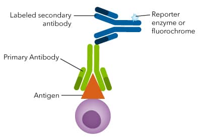
A secondary antibody is an antibody that is used to bind the immunoglobulin (IgG) domain of the primary antibody. The secondary antibody will be specific to the primary antibody’s species and isotype. A secondary antibody is usually conjugated with a molecule that allows for the antibody to be detected. The molecules can be chromogenic or fluorescent.

Primary and Secondary Antibody


How To Select A Secondary Antibody

When selecting a secondary antibody, it should be chosen based off the primary antibody and have the appropriate conjugate for the application it is being used in. Since the secondary antibody is used to detect the primary antibody, the secondary antibody should detect the host species of the primary antibody. For instance, if the primary antibody is a goat anti-human (raised in a goat host, recognizing a human protein) then the secondary antibody needs to be specific to goat IgG. A common example would be a donkey anti-goat IgG (raised in a donkey host to detect goat IgG).

Detection of ACE-2 Antibody in Human PBMC by Flow Cytometry

Detection of Mouse CD31/PECAM‑1 by Western Blot. Western blot shows lysates of bEnd.3 mouse endothelioma cell line. PVDF membrane was probed with 0.5 µg/mL of Goat Anti-Mouse/Rat CD31/PECAM-1 Antigen Affinity-purified Polyclonal Antibody (Catalog # AF3628) followed by HRP-conjugated Anti-Goat IgG Secondary Antibody (Catalog # HAF017). A specific band was detected for CD31/PECAM-1 at approximately 130 kDa (as indicated). This experiment was conducted under reducing conditions and using our Western blot Buffer Group 1.


VisUCyte™ HRP Polymer

VisUCyte™ HRP Polymer Antibodies are a biotin-free detection reagent that removes the need to perform additional quenching of endogenous biotin and avidin present in some tissues. By removing the need for need for additional blocking and incubation steps, researchers can achieve specific staining in cells and tissue much faster than the conventional avidin-biotin-HRP procedure. VisUCyte HRP polymer also provides higher sensitivity allowing researchers to use 2-3 times less primary antibody when compared to the conventional avidin-biotin-HRP protocols.

Learn About VisUCyte HRP Polymer

For added convenience, we also offer Cell and Tissue Staining Kits that include secondary antibodies, blocking reagents, streptavidin HRP, and a chromogen reagent.

Browse Cell and Tissue Staining Kits

Secondary antibodies conjugated to VisUCyte HRP Polymer


NorthernLights™ Secondary Antibodies and Streptavidin Conjugates

NorthernLights™ fluorescent secondary antibodies and streptavidin conjugates are bright and resistant to photobleaching. These conjugates are great options that work well in Flow cytometry and fluorescence microscopy. Their spectral properties are similar to several widely used fluorochromes and are suitable for use with common filter sets and lasers. These secondary antibodies are available with three distinct excitation and emission wavelengths, making them an optimal option for multi-color experiments. NorthernLights secondary antibodies are stable when exposed to alcohols and xylene, as well as DPX mounting medium making these fluorescent conjugates ideal for fluorescent microscopy.

View all NorthernLight Products

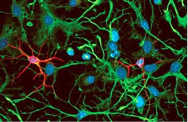
Rat cortical stem cell staining using NortherLights secondary antibodies

Rat cortical stem cell differentiation was monitored using multi-color immunocytochemistry. Neural progenitors were labeled with Goat anti-Rat Nestin Polyclonal Antibody (Catalog # AF2736) and stained with Donkey anti-Goat NorthernLights-493 Secondary Antibody (Catalog # NL003; green). Differentiated neurons were labeled with Mouse Anti-Neuron-specific beta -III Tubulin Monoclonal Antibody (Catalog # MAB1195) and stained using Donkey anti-Mouse NorthernLights-557 Secondary Antibody (Catalog # NL007; red). Nuclei were stained with DAPI (blue).