Human Arginase 1/ARG1 Antibody Summary
Met1-Lys322
Accession # P05089
*Small pack size (-SP) is supplied either lyophilized or as a 0.2 µm filtered solution in PBS.
Applications
Please Note: Optimal dilutions should be determined by each laboratory for each application. General Protocols are available in the Technical Information section on our website.
Scientific Data
 View Larger
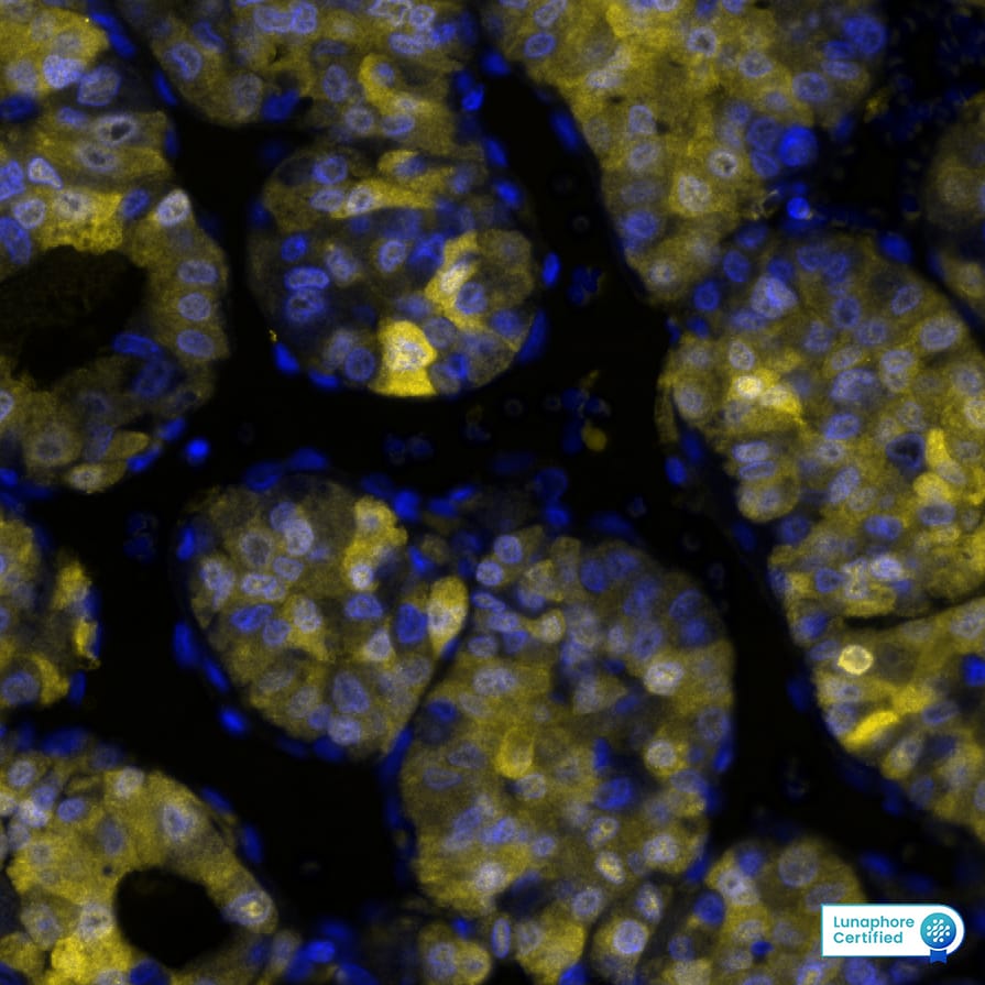
Detection of Arginase1 in Human Liver Tumor via seqIF™ staining on COMET™ Arginase 1 was detected in immersion fixed paraffin-embedded sections of human Liver Tumor using Mouse Anti-Human Arginase 1, Monoclonal Antibody (Catalog # MAB5868) at 3ug/mL at 37° Celsius for 4 minutes. Before incubation with the primary antibody, tissue underwent an all-in-one dewaxing and antigen retrieval preprocessing using PreTreatment Module (PT Module) and Dewax and HIER Buffer H (pH 9; Epredia Catalog # TA-999-DHBH). Tissue was stained using the Alexa Fluor™ 647 Goat anti-Mouse IgG Secondary Antibody at 1:200 at 37 ° Celsius for 2 minutes. (Yellow; Lunaphore Catalog # DR647MS) and counterstained with DAPI (blue; Lunaphore Catalog # DR100). Specific staining was localized to the cytoplasm and nucleus. Protocol available in COMET™ Panel Builder.
 View Larger
Detection of Human Arginase 1/ARG1 by Western Blot. Western blot shows lysates of human liver tissue. PVDF Membrane was probed with 1 µg/mL of Human Arginase 1/ARG1 Monoclonal Antibody (Catalog # MAB5868) followed by HRP-conjugated Anti-Mouse IgG Secondary Antibody (Catalog # HAF007). A specific band was detected for Arginase 1/ARG1 at approximately 40 kDa (as indicated). This experiment was conducted under reducing conditions and using Immunoblot Buffer Group 1.
 View Larger
Detection of Human Arginase 1/ARG1 by Simple WesternTM. Simple Western lane view shows lysates of human liver tissue, loaded at 0.2 mg/mL. A specific band was detected for Arginase 1/ARG1 at approximately 42 kDa (as indicated) using 20 µg/mL of Mouse Anti-Human Arginase 1/ARG1 Monoclonal Antibody (Catalog # MAB5868). This experiment was conducted under reducing conditions and using the 12-230 kDa separation system.*Non-specific interaction with the 230kDa Simple Western standard may be seen with this antibody.
 View Larger
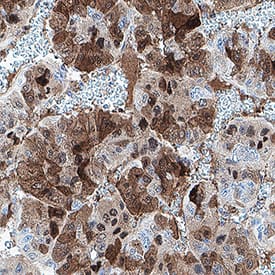
Detection of Arginase 1/ARG1 in Human Liver Cancer. Arginase 1/ARG1 was detected in immersion fixed paraffin-embedded sections of human liver cancer using Mouse Anti-Human Arginase 1/ARG1 Monoclonal Antibody (Catalog # MAB5868) at 1 µg/ml overnight at 4 °C. Before incubation with the primary antibody, tissue was subjected to heat-induced epitope retrieval using VisUCyte Antigen Retrieval Reagent-Basic (Catalog # VCTS021). Tissue was stained using the HRP-conjugated Anti-Mouse IgG Secondary Antibody (Catalog # HAF007) and counterstained with hematoxylin (blue). Specific staining was localized to the nucleus and cytoplasm. View our protocol for Chromogenic IHC Staining of Paraffin-embedded Tissue Sections.
 View Larger
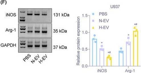
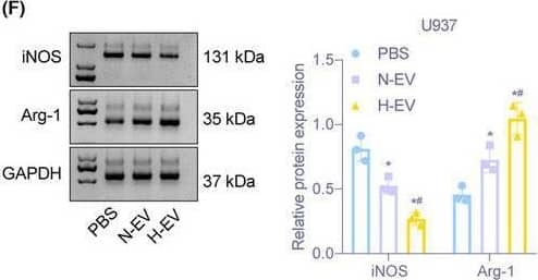
Detection of Arginase 1/ARG1 by Western Blot Hypoxic glioma cell‐derived EVs promote M2 macrophage polarization. (A) Images of EVs observed under a TEM (Scale bar = 100 nm). (B) Diameter of EVs detected by dynamic light scattering. (C) The expression of CD9, CD63, TSG101, and calnexin on the surface of EVs determined by Immunoblotting. (D) Detection of the uptake of EVs from normoxic and hypoxic glioma cells by macrophages U937 through immunofluorescence (400×). (E) The mRNA expression of iNOS, TNF‐ alpha, Arg‐1, and IL‐10 assessed by RT‐qPCR after EV treatment. (F) Immunoblotting of protein expression of iNOS and Arg‐1 in macrophages U937 after EV treatment. (G) The expression of TNF‐ alpha and IL‐10 in supernatant of macrophages U937 evaluated by ELISA assay. (H) The proportion of CD11b+CD163+ cells in U937 cells examined by flow cytometry. *p < 0.05 versus macrophages treated with PBS. #p < 0.05 versus macrophages treated with normal oxygen‐induced glioma cell‐derived EVs. The experiment was repeated three times Image collected and cropped by CiteAb from the following open publication (https://pubmed.ncbi.nlm.nih.gov/36052751), licensed under a CC-BY license. Not internally tested by R&D Systems.
Reconstitution Calculator
Preparation and Storage
- 12 months from date of receipt, -20 to -70 °C as supplied.
- 1 month, 2 to 8 °C under sterile conditions after reconstitution.
- 6 months, -20 to -70 °C under sterile conditions after reconstitution.
Background: Arginase 1/ARG1
Arginase 1 (ARG1) is a 35‑40 kDa member of the arginase family of enzymes. It is expressed in multiple cell types, including erythrocytes, hepatocytes, neutrophils, smooth muscle and macrophages. ARG1 demonstrates two distinct functions: in the hepatocyte cytoplasm, it catalyzes the conversion of arginine to ornithine and urea, while in multiple cells, it degrades arginine, thus indirectly downregulating NO synthase (NOS) activity by depriving this enzyme of its substrate. Human AGR1 is 322 amino acids (aa) in length. Its enzyme region comprises aa 9‑309 and contains two Mn atoms. ARG1 is moderately active as a monomer, but highly active as a 105 kDa homotrimer. Trimerization is promoted by nitrosylation of Cys303, creating a regulatory feedback loop with NOS. There are two isoform variants, one that shows an eight aa insertion after Gln43, and another that shows a deletion of aa 204‑289. Full-length human ARG1 shares 87% aa identity with mouse and rat ARG1.
Product Datasheets
Citation for Human Arginase 1/ARG1 Antibody
R&D Systems personnel manually curate a database that contains references using R&D Systems products. The data collected includes not only links to publications in PubMed, but also provides information about sample types, species, and experimental conditions.
1 Citation: Showing 1 - 1
-
Hypoxic glioma‐derived extracellular vesicles harboring MicroRNA‐10b‐5p enhance M2 polarization of macrophages to promote the development of glioma
Authors: Bingzhen Li, Cheng Yang, Zhanpeng Zhu, Hao Chen, Bin Qi
CNS Neuroscience & Therapeutics
FAQs
No product specific FAQs exist for this product, however you may
View all Antibody FAQsReviews for Human Arginase 1/ARG1 Antibody
Average Rating: 4.5 (Based on 2 Reviews)
Have you used Human Arginase 1/ARG1 Antibody?
Submit a review and receive an Amazon gift card.
$25/€18/£15/$25CAN/¥75 Yuan/¥2500 Yen for a review with an image
$10/€7/£6/$10 CAD/¥70 Yuan/¥1110 Yen for a review without an image
Filter by:

