Human B7-H3 Antibody Summary
Leu29-Pro245
Accession # Q5ZPR3
Applications
Please Note: Optimal dilutions should be determined by each laboratory for each application. General Protocols are available in the Technical Information section on our website.
Scientific Data
 View Larger
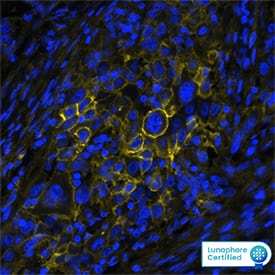
Detection of B7-H3 in Human Lung Cancer via Multiplex Immunofluorescence staining on COMET™ B7-H3 was detected in immersion fixed paraffin-embedded sections of human lung cancer using Mouse Anti-Human B7-H3 Monoclonal Antibody (Catalog # MAB11611) at 20ug/mL at 37 ° Celsius for 4 minutes. Before incubation with the primary antibody, tissue underwent an all-in-one dewaxing and antigen retrieval preprocessing using PreTreatment Module (PT Module) and Dewax and HIER Buffer H (pH 9). Tissue was stained using the Alexa Fluor™ 647 Goat anti-Mouse IgG Secondary Antibody at 1:200 at 37 ° Celsius for 2 minutes. (Yellow; Lunaphore Catalog # DR647MS) and counterstained with DAPI (blue; Lunaphore Catalog # DR100). Specific staining was localized to the membrane. Protocol available in COMET™ Panel Builder.
 View Larger
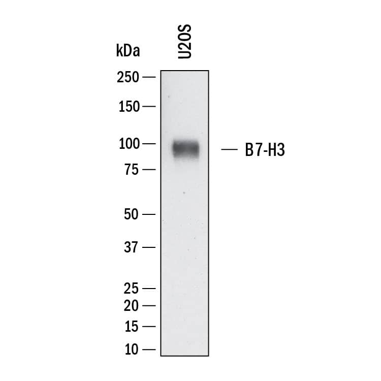
Detection of Human B7‑H3 by Western Blot. Western Blot shows lysates of U2OS human osteosarcoma cell line. PVDF membrane was probed with 1 µg/ml of Mouse Anti-Human B7‑H3 Monoclonal Antibody (Catalog # MAB11611) followed by HRP-conjugated Anti-Mouse IgG Secondary Antibody (Catalog # HAF018). A specific band was detected for B7‑H3 at approximately 90 kDa (as indicated). This experiment was conducted under reducing conditions and using Western Blot Buffer Group 1.
 View Larger
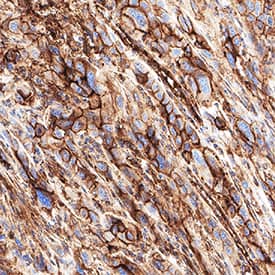
Detection of B7‑H3 in Human Lung Cancer. B7‑H3 was detected in immersion fixed paraffin-embedded sections of human lung cancer using Mouse Anti-Human B7‑H3 Monoclonal Antibody (Catalog # MAB11611) at 5 µg/ml for 1 hour at room temperature followed by incubation with the Anti-Mouse IgG VisUCyte™ HRP Polymer Antibody (Catalog # VC001). Before incubation with the primary antibody, tissue was subjected to heat-induced epitope retrieval using VisUCyte Antigen Retrieval Reagent-Basic (Catalog # VCTS021). Tissue was stained using DAB (brown) and counterstained with hematoxylin (blue). Specific staining was localized to the cell membrane. View our protocol for IHC Staining with VisUCyte HRP Polymer Detection Reagents.
 View Larger
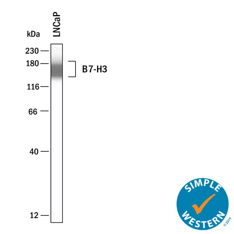
Detection of Human B7‑H3 by Simple WesternTM. Simple Western shows lysates of LNCaP human prostate cancer cell line, loaded at 0.2 mg/ml. A specific band was detected for B7‑H3 at approximately 161 kDa (as indicated) using 20 µg/mL of Mouse Anti-Human B7‑H3 Monoclonal Antibody (Catalog # MAB11611). This experiment was conducted under reducing conditions and using the 12‑230 kDa separation system.
Reconstitution Calculator
Preparation and Storage
- 12 months from date of receipt, -20 to -70 °C as supplied.
- 1 month, 2 to 8 °C under sterile conditions after reconstitution.
- 6 months, -20 to -70 °C under sterile conditions after reconstitution.
Background: B7-H3
Human B7 homolog 3 (B7-H3) is a member of the B7 family of immune proteins that provide signals for regulating immune responses (1‑3). Other family members include B7-1, B7-2, B7-H2, PD-L1 (B7-H1), and PD-L2. B7 proteins are immunoglobulin (Ig) superfamily members with extracellular Ig-V-like and Ig-C-like domains and short cytoplasmic domains. Among the family members, they share about 20‑40% amino acid (aa) sequence identity. The cloned human B7-H3 cDNA encodes a 316 aa type I membrane precursor protein with a putative 28 aa signal peptide, a 217 aa extracellular region containing one V-like and one C-like Ig domain, a transmembrane region, and a 45 aa cytoplasmic domain. An isoform of human B7-H3 containing a four-Ig-like domain extracellular region has also been identified. Human B7-H3 is not expressed on resting B cells, T cells, monocytes or dendritic cells, but is induced on dendritic cells and monocytes by inflammatory cytokines. B7-H3 expression is also detected on various normal tissues and in some tumor cell lines. Human B7-H3 does not bind any known members of the CD28 family of immunoreceptors. However, B7-H3 has been shown to bind an unidentified counter-receptor on activated T cells to costimulate the proliferation of CD4+ or CD8+ T cells. B7-H3 has also been found to enhance the induction of primary cytotoxic T lymphocytes and stimulate IFN-gamma production (1‑3).
- Chapoval, A.I. et al. (2001) Nat. Immunol. 2:269.
- Sharpe, A.H. and G.J. Freeman (2002) Nat. Rev. Immunol. 2:116.
- Coyle, A. and J. Gutierrez-Ramos (2001) Nat. Immunol. 2:203.
Product Datasheets
FAQs
No product specific FAQs exist for this product, however you may
View all Antibody FAQsReviews for Human B7-H3 Antibody
There are currently no reviews for this product. Be the first to review Human B7-H3 Antibody and earn rewards!
Have you used Human B7-H3 Antibody?
Submit a review and receive an Amazon gift card.
$25/€18/£15/$25CAN/¥75 Yuan/¥2500 Yen for a review with an image
$10/€7/£6/$10 CAD/¥70 Yuan/¥1110 Yen for a review without an image

