Human BAK Antibody Summary
AAPADPEMVTLPLQPSSTMGC
Accession # Q16611
Applications
Please Note: Optimal dilutions should be determined by each laboratory for each application. General Protocols are available in the Technical Information section on our website.
Scientific Data
 View Larger
Detection of Human BAK by Western Blot. Western blot shows lysates of THP-1 human acute monocytic leukemia cell line and HEK293 human embryonic kidney cell line. PVDF membrane was probed with 1-4 µg/mL of Human BAK Antigen Affinity-purified Polyclonal Antibody (Catalog # AF816) followed by HRP-conjugated Anti-Rabbit IgG Secondary Antibody (HAF008). A specific band was detected for BAK at approximately 26 kDa (as indicated). This experiment was conducted under reducing conditions and using Immunoblot Buffer Group 2.
 View Larger
BAK in HEK293 Human Cell Line. BAK was detected in immersion fixed HEK293 human embryonic kidney cell line using Rabbit Anti-Human BAK Antigen Affinity-purified Polyclonal Antibody (Catalog # AF816) at 15 µg/mL for 3 hours at room temperature. Cells were stained using the NorthernLights™ 557-conjugated Anti-Rabbit IgG Secondary Antibody (red; NL004) and counterstained with DAPI (blue). Specific staining was localized to cytoplasm. View our protocol for Fluorescent ICC Staining of Cells on Coverslips.
 View Larger
Detection of BAK in Human Colon. BAK was detected in immersion fixed paraffin-embedded sections of Human Colon using Rabbit Anti-Human BAK Antigen Affinity-purified Polyclonal Antibody (Catalog # AF816) at 5 µg/mL for 1 hour at room temperature followed by incubation with the Anti-Rabbit IgG VisUCyte™ HRP Polymer Antibody (Catalog # VC003). Before incubation with the primary antibody, tissue was subjected to heat-induced epitope retrieval using VisUCyte Antigen Retrieval Reagent-Basic (Catalog # VCTS021). Tissue was stained using DAB (brown) and counterstained with hematoxylin (blue). Specific staining was localized to cell membrane in epithelial cells in mucosal glands. View our protocol for IHC Staining with VisUCyte HRP Polymer Detection Reagents.
 View Larger
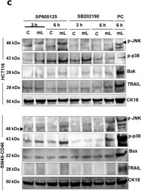
Detection of BAK by Western Blot Role of TRAF3 & pro-apoptotic p38 & JNK kinases in mCD40L-mediated intrinsic & extrinsic cell death signalling pathways. (c) Untreated (‘C’) & mCD40L-treated (‘mL’) HCT116 & SW480-CD40 in the presence of 5µM JNK inhibitor SP600125 or p38 inhibitor SB202190 used to detect phosphorylated JNK (p-JNK) & p38 (p-p38), pro-apoptotic Bak or Bax, & TRAIL protein by immunoblotting at 3 h & 6 h post CD40 ligation. Lysates from HCT116 & SW480-CD40 cell cultures treated with mCD40L for 6 h in the absence of inhibitor included (denoted as positive control, ‘PC’) per experiment, respectively. Equal loading for human epithelial cell lysate was confirmed by CK18 detection in all experiments. Image collected & cropped by CiteAb from the following open publication (https://pubmed.ncbi.nlm.nih.gov/36291141), licensed under a CC-BY license. Not internally tested by R&D Systems.
 View Larger
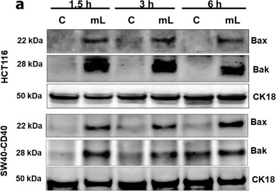
Detection of BAK by Western Blot Rapid induction of the mitochondrial (intrinsic) apoptotic pathway and role of caspase activation in cell death. (a) Expression of Bax and Bak proteins was detected in controls (‘C’) versus mCD40L-treated (‘mL’) HCT116 and SW480-CD40 cells by immunoblotting at the indicated time points. Equal loading for human epithelial cell lysate was confirmed by CK18 detection. (b) Control (‘C’) and mCD40L-treated (‘mL’) HCT116 and SW480-CD40 cells were used to prepare cytoplasmic (‘Cyto’) and mitochondrial (‘Mito’) sub-cellular fractions for the detection of cytochrome c (Cyto c) protein by immunoblotting at 6 h post CD40 ligation. Detection of Bcl-2 and GAPDH proteins was employed to confirm sub-cellular fractionation (mitochondrial and cytoplasmic, respectively). (c) HCT116 cells were treated with mCD40L in the absence (vehicle control–denoted ‘Control’) or presence of 100 µM inhibitor of caspase-8 (z-IETD-FMK), caspase-9 (z-LEHD-FMK), caspase-10 (z-AEVD-FMK) or pan-caspase inhibitor (z-VAD-FMK). Cell death was detected 24 h later using the CytoTox-Glo assay. Results are presented as Cell death Fold increase in background-corrected RLU readings relative to control (mCD40L treatment versus controls) and are representative of 3 independent experiments. Bars show mean fold change (comparing caspase inhibitor-treated versus vehicle control cultures) for 5–6 technical replicates ± SEM. NS denotes non-significance (p > 0.05) and *** p < 0.001. Image collected and cropped by CiteAb from the following open publication (https://pubmed.ncbi.nlm.nih.gov/36291141), licensed under a CC-BY license. Not internally tested by R&D Systems.
Reconstitution Calculator
Preparation and Storage
- 12 months from date of receipt, -20 to -70 °C as supplied.
- 1 month, 2 to 8 °C under sterile conditions after reconstitution.
- 6 months, -20 to -70 °C under sterile conditions after reconstitution.
Background: BAK
BAK (Bcl-2 homologous antagonist/killer; also BAK1) is a 25‑30 kDa member of the BCL-2 family of proteins. It is widely expressed, and participates in the apoptotic cycle. BAK is an outer mitochondrial membrane protein that is inactive as a Zn-dependent homodimer. Upon activation by p53 or tBID, BAK oligomerizes, creating a pore in the mitochondrial membrane and allowing for cytochrome C release. Human BAK is 211 amino acids (aa) in length and contains three BCL-2 homology domains (aa 74‑88, 117‑136 and 169‑184), a Zn-binding region (aa 160‑166) and a C-terminal transmembrane segment (aa 188‑205). Amino acids 67‑94 mediate oligomerization of BAK. There are two potential isoform variants; one shows an alternate start site at Met96, while a second shows a deletion of aa 46‑66. Over amino acids 53-72, human BAK shares 55% aa identity with mouse BAK.
Product Datasheets
Citations for Human BAK Antibody
R&D Systems personnel manually curate a database that contains references using R&D Systems products. The data collected includes not only links to publications in PubMed, but also provides information about sample types, species, and experimental conditions.
5
Citations: Showing 1 - 5
Filter your results:
Filter by:
-
TRAF3/p38-JNK Signalling Crosstalk with Intracellular-TRAIL/Caspase-10-Induced Apoptosis Accelerates ROS-Driven Cancer Cell-Specific Death by CD40
Authors: K Ibraheem, AMA Yhmed, MM Nasef, NT Georgopoul
Cells, 2022-10-18;11(20):.
Species: Human
Sample Types: Cell Lysates
Applications: Western Blot -
CD40 induces renal cell carcinoma-specific differential regulation of TRAF proteins, ASK1 activation and JNK/p38-mediated, ROS-dependent mitochondrial apoptosis
Authors: Khalidah Ibraheem, Albashir M. A. Yhmed, Tahir Qayyum, Nicolas P. Bryan, Nikolaos T. Georgopoulos
Cell Death Discovery
-
A redox state-dictated signalling pathway deciphers the malignant cell specificity of CD40-mediated apoptosis
Oncogene, 2016-11-21;0(0):.
Species: Human
Sample Types: Cell Lysates
Applications: Western Blot -
Human liver sinusoidal endothelial cells induce apoptosis in activated T cells: a role in tolerance induction.
Authors: Karrar A, Broome U, Uzunel M, Qureshi AR, Sumitran-Holgersson S
Gut, 2006-07-13;56(2):243-52.
Species: Human
Sample Types: Cell Lysates
Applications: Western Blot -
Apoptotic signaling pathways induced by nitric oxide in human lymphoblastoid cells expressing wild-type or mutant p53.
Authors: Li CQ, Robles AI, Hanigan CL, Hofseth LJ, Trudel LJ, Harris CC, Wogan GN
Cancer Res., 2004-05-01;64(9):3022-9.
Species: Human
Sample Types: Cell Lysates
Applications: Western Blot
FAQs
No product specific FAQs exist for this product, however you may
View all Antibody FAQsReviews for Human BAK Antibody
There are currently no reviews for this product. Be the first to review Human BAK Antibody and earn rewards!
Have you used Human BAK Antibody?
Submit a review and receive an Amazon gift card.
$25/€18/£15/$25CAN/¥75 Yuan/¥2500 Yen for a review with an image
$10/€7/£6/$10 CAD/¥70 Yuan/¥1110 Yen for a review without an image


