Human Caspase-1 Antibody Summary
Asn120-Asp297
Accession # P29466
Applications
This antibody functions as an ELISA detection antibody when paired with Mouse Anti-Human Caspase‑1 Monoclonal Antibody (Catalog # MAB62151).
This product is intended for assay development on various assay platforms requiring antibody pairs. We recommend the Human Caspase-1/ICE Quantikine ELISA Kit (Catalog # DCA100) for a complete optimized ELISA.
Please Note: Optimal dilutions should be determined by each laboratory for each application. General Protocols are available in the Technical Information section on our website.
Scientific Data
 View Larger
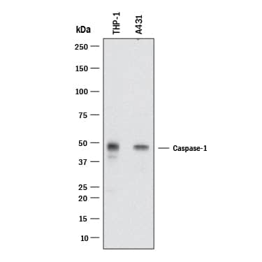
Detection of Human Caspase‑1 by Western Blot. Western blot shows lysates of THP‑1 human acute monocytic leukemia cell line and A431 human epithelial carcinoma cell line. PVDF membrane was probed with 2 µg/mL of Mouse Anti-Human Caspase‑1 Monoclonal Antibody (Catalog # MAB62153) followed by HRP-conjugated Anti-Mouse IgG Secondary Antibody (HAF018). A specific band was detected for Caspase‑1 at approximately 48 kDa (as indicated). This experiment was conducted under reducing conditions and using Western Blot Buffer Group 1.
 View Larger
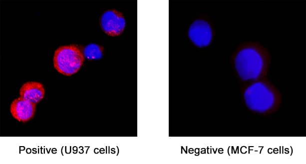
Caspase‑1 in U937 Human Cell Line. Caspase‑1 was detected in immersion fixed U937 human histiocytic lymphoma cell line (positive) and MCF‑7 human breast cancer cell line (negative control) using Mouse Anti-Human Caspase‑1 Monoclonal Antibody (Catalog # MAB62153) at 8 µg/mL for 3 hours at room temperature. Cells were stained using the NorthernLights™ 557-conjugated Anti-Mouse IgG Secondary Antibody (red; NL007) and counterstained with DAPI (blue). Specific staining was localized to cell surface and cytoplasm. Staining was performed using our protocol for Fluorescent ICC Staining of Non-adherent Cells.
 View Larger
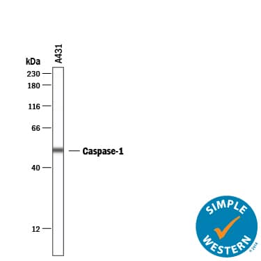
Detection of Human Caspase‑1 by Simple WesternTM. Simple Western lane view shows lysates of A431 human epithelial carcinoma cell line, loaded at 0.2 mg/mL. A specific band was detected for Caspase‑1 at approximately 52 kDa (as indicated) using 20 µg/mL of Mouse Anti-Human Caspase‑1 Monoclonal Antibody (Catalog # MAB62153). This experiment was conducted under reducing conditions and using the 12-230 kDa separation system.
 View Larger
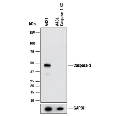
Western Blot Shows Human Caspase‑1 Specificity by Using Knockout Cell Line. Western blot shows lysates of A431 human epithelial carcinoma parental cell line and Caspase‑1 knockout A431 cell line (KO). PVDF membrane was probed with 2 µg/mL of Mouse Anti-Human Caspase‑1 Monoclonal Antibody (Catalog # MAB62153) followed by HRP-conjugated Anti-Mouse IgG Secondary Antibody (HAF018). A specific band was detected for Caspase‑1 at approximately 48 kDa (as indicated) in the parental A431 cell line, but is not detectable in knockout A431 cell line. GAPDH (2275-PC-100) is shown as a loading control. This experiment was conducted under reducing conditions and using Western Blot Buffer Group 1.
 View Larger
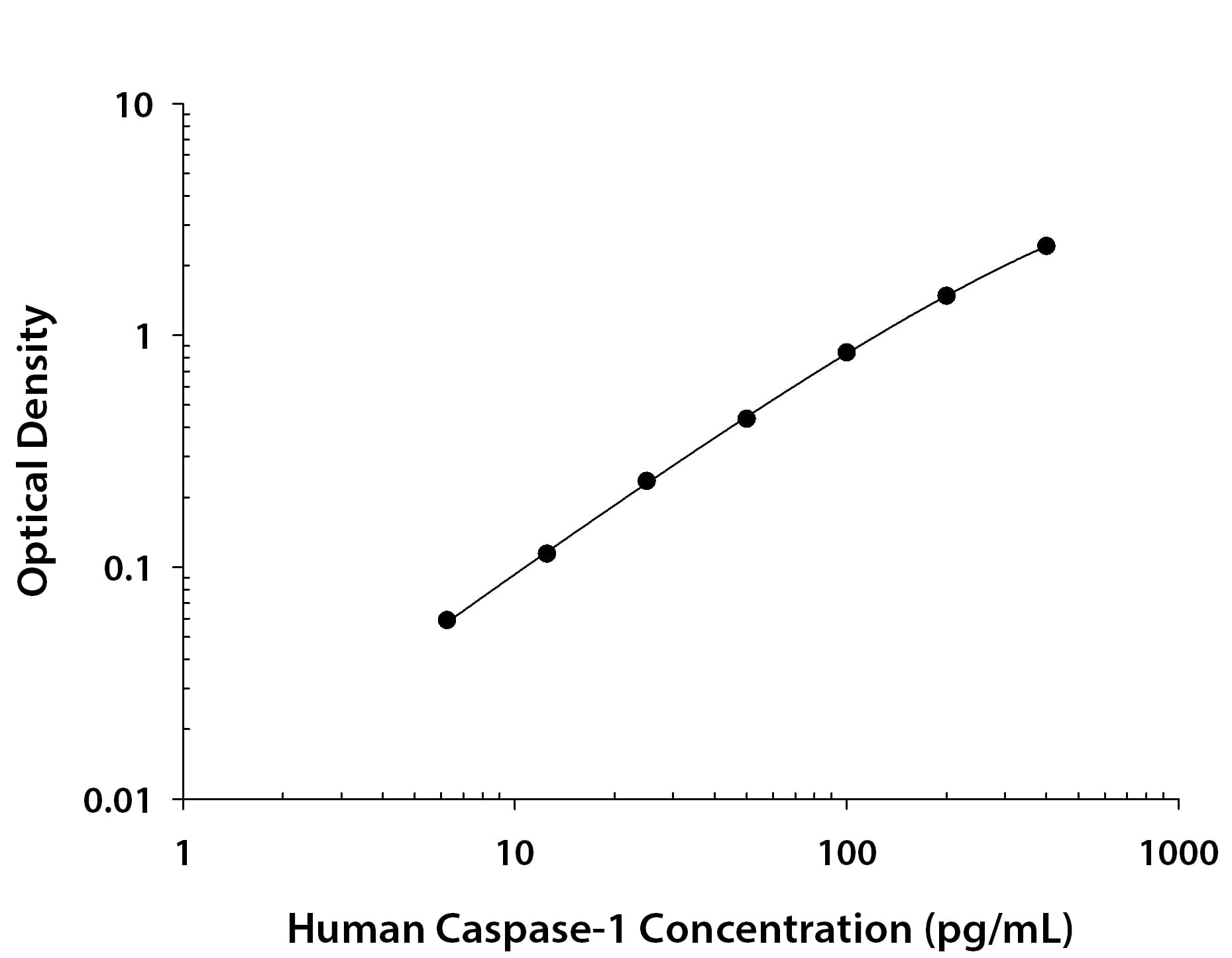
Human Caspase‑1 ELISA Standard Curve. Recombinant Human Caspase‑1 protein was serially diluted 2-fold and captured by Mouse Anti-Human Caspase‑1 Monoclonal Antibody (MAB62151) coated on a Clear Polystyrene Microplate (DY990). Mouse Anti-Human Caspase‑1 Monoclonal Antibody (Catalog # MAB62153) was biotinylated and incubated with the protein captured on the plate. Detection of the standard curve was achieved by incubating Streptavidin-HRP (DY998) followed by Substrate Solution (DY999) and stopping the enzymatic reaction with Stop Solution (DY994).
Reconstitution Calculator
Preparation and Storage
- 12 months from date of receipt, -20 to -70 °C as supplied.
- 1 month, 2 to 8 °C under sterile conditions after reconstitution.
- 6 months, -20 to -70 °C under sterile conditions after reconstitution.
Background: Caspase-1
Caspase-1, also known as IL-1 beta -Converting Enzyme (ICE), is an aspartic protease that plays a key role in the inflammatory response and apoptosis. Caspase-1 precursor (about 50kDa) can be cleaved and the active enzyme consists of a complex of two 20 kDa (aa 120-297) and two 10 kDa (aa 317-404) subunits which associate following cleavage of inactive precursors. Caspase-1 is required for proteolytic cleavage of the IL-1 beta precursor to form the active proinflammatory cytokine. Alternate splicing generates several additional Caspase-1 isoforms with deletions in the propeptide regions or also in the mature subunits. Within the large subunit, human Caspase 1 shares 61% aa sequence identity with mouse and rat Caspase-1.
Product Datasheets
FAQs
No product specific FAQs exist for this product, however you may
View all Antibody FAQsReviews for Human Caspase-1 Antibody
Average Rating: 5 (Based on 1 Review)
Have you used Human Caspase-1 Antibody?
Submit a review and receive an Amazon gift card.
$25/€18/£15/$25CAN/¥75 Yuan/¥2500 Yen for a review with an image
$10/€7/£6/$10 CAD/¥70 Yuan/¥1110 Yen for a review without an image
Filter by:


