Human CD14 Antibody Summary
Applications
Please Note: Optimal dilutions should be determined by each laboratory for each application. General Protocols are available in the Technical Information section on our website.
Scientific Data
 View Larger
Detection of Human CD14 by Western Blot. Western blot shows lysates of THP‑1 human acute monocytic leukemia cell line untreated (-) or treated (+) with 200 nM of PMA for 24 hours and 10 ug/mL of LPS for 3 hours. PVDF membrane was probed with 1 µg/mL of Sheep Anti-Human CD14 Polyclonal Antibody (Catalog # AB383) followed by HRP-conjugated Anti-Sheep IgG Secondary Antibody (HAF016). A specific band was detected for CD14 at approximately 55 kDa (as indicated). This experiment was conducted under non-reducing conditions and using Western Blot Buffer Group 1.
 View Larger
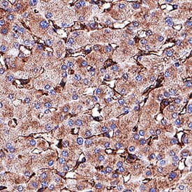
Detection of CD14 in Human Liver. CD14 was detected in immersion fixed paraffin-embedded sections of Human Liver using Sheep Anti-Human CD14 Polyclonal Antibody (Catalog # AB383) at 3 µg/mL for 1 hour at room temperature followed by incubation with the Anti-Sheep IgG VisUCyte™ HRP Polymer Antibody (Catalog # VC006). Before incubation with the primary antibody, tissue was subjected to heat-induced epitope retrieval using VisUCyte Antigen Retrieval Reagent-Basic (Catalog # VCTS021). Tissue was stained using DAB (brown) and counterstained with hematoxylin (blue). Specific staining was localized to sinusoids. View our protocol for IHC Staining with VisUCyte HRP Polymer Detection Reagents.
 View Larger
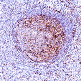
Detection of CD14 in Human Tonsil. CD14 was detected in immersion fixed paraffin-embedded sections of Human Tonsil using Sheep Anti-Human CD14 Polyclonal Antibody (Catalog # AB383) at 3 µg/mL for 1 hour at room temperature followed by incubation with the Anti-Sheep IgG VisUCyte™ HRP Polymer Antibody (Catalog # VC006). Before incubation with the primary antibody, tissue was subjected to heat-induced epitope retrieval using VisUCyte Antigen Retrieval Reagent-Basic (Catalog # VCTS021). Tissue was stained using DAB (brown) and counterstained with hematoxylin (blue). Specific staining was localized to lymphocytes in germinal center. View our protocol for IHC Staining with VisUCyte HRP Polymer Detection Reagents.
Preparation and Storage
- 12 months from date of receipt, -20 to -70 °C as supplied.
- 1 month, 2 to 8 °C under sterile conditions after reconstitution.
- 6 months, -20 to -70 °C under sterile conditions after reconstitution.
Background: CD14
CD14 is a 55 kDa cell surface glycoprotein that is preferentially expressed on monocytes/macrophages. The human CD14 cDNA encodes a 375 amino acid (aa) residue precursor protein with a 19 aa signal peptide and a C-terminal hydrophobic region characteristic for glycosylphosphatidyinositol (GPI)-anchored proteins. Human CD14 has four potential N-linked glycosylation sites and also bears O-linked carbohydrates. The amino acid sequence of human CD14 is approximately 65% identical with the mouse, rat, rabbit, and bovine proteins. CD14 is a pattern recognition receptor that binds lipopolysaccharides (LPS) and a variety of ligands derived from different microbial sources. The binding of CD14 with LPS is catalyzed by LPS-binding protein (LBP). The toll-like-receptors have also been implicated in the transduction of CD14-LPS signals. Similar to other GPI-anchored proteins, soluble CD14 can be released from the cell surface by phosphatidyinositol-specific phospholipase C. Soluble CD14 has been detected in serum and body fluids. High concentrations of soluble CD14 have been shown to inhibit LPS-mediated responses. However, soluble CD14 can also potentiate LPS response in cells that do not express cell surface CD14.
- Wright, S.D. et al. (1990) Science 249:1431.
- Pugin, J. et al. (1993) Proc. Natl. Acad. Sci. USA 90:2744.
- Beutler, B. (2000) Current Opinion in Immunology 12:20.
- Stelter, F. (2000) Chem. Immunol. 74:25.
Product Datasheets
Citations for Human CD14 Antibody
R&D Systems personnel manually curate a database that contains references using R&D Systems products. The data collected includes not only links to publications in PubMed, but also provides information about sample types, species, and experimental conditions.
2
Citations: Showing 1 - 2
Filter your results:
Filter by:
-
Dermal CD14(+) Dendritic Cell and Macrophage Infection by Dengue Virus Is Stimulated by Interleukin-4.
Authors: Schaeffer E, Flacher V, Papageorgiou V, Decossas M, Fauny J, Kramer M, Mueller C
J Invest Dermatol, 2014-12-18;135(7):1743-51.
Species: Human
Sample Types: Whole Cells
Applications: Flow Cytometry, IHC -
Induction of keratinocyte growth factor 1 Expression by lipopolysaccharide is regulated by CD-14 and toll-like receptors 2 and 4.
Authors: Putnins EE, Sanaie AR, Wu Q
Infect. Immun., 2002-12-01;70(12):6541-8.
Species: Human
Sample Types: Cell Lysates, Whole Cells
Applications: Neutralization, Western Blot
FAQs
No product specific FAQs exist for this product, however you may
View all Antibody FAQsReviews for Human CD14 Antibody
There are currently no reviews for this product. Be the first to review Human CD14 Antibody and earn rewards!
Have you used Human CD14 Antibody?
Submit a review and receive an Amazon gift card.
$25/€18/£15/$25CAN/¥75 Yuan/¥2500 Yen for a review with an image
$10/€7/£6/$10 CAD/¥70 Yuan/¥1110 Yen for a review without an image


