Carcinoembryonic antigen (CEA)-related cell adhesion molecule 1 (CEACAM-1; also BGP) is a 160 kDa member of the CEACAM branch of the CEA gene family of the immunoglobulin superfamily (1-3). It is one of seven human CEACAM subfamily genes that are essentially divided equally between type I transmembrane proteins (CEACAM-1, 3, and 4) and GPI-linked molecules (CEACAM-5-8). There is no CEACAM-2 in human. The gene for human CEACAM-1 codes for a 526 amino acid (aa) type I transmembrane protein that contains a 34 aa signal sequence, a 394 aa extracellular domain (ECD), a 24 aa transmembrane segment, and a 74 aa cytoplasmic region (4, 5). The ECD contains one N-terminal V-type Ig-like domain, followed by three C2-type Ig-like domains. It shows considerable glycosylation, including high mannose residues and (sialyl) LewisX (1). The cytoplasmic region shows one ITIM motif and a calmodulin binding site (1-3). In addition to the full length form, ten alternate splice forms have been reported (1, 4, 6-8). There are three soluble and seven transmembrane isoforms, with variations occurring in both the ECD and cytoplasmic region. All ten alternate splice forms contain the V-type Ig-like domain (aa’s 35-142). The three soluble forms also contain the first two C2-type Ig-like domains (aa’s 145-317), with differences coming in the third C2-type Ig-like domain (6). The seven transmembrane isoforms are highly divergent. Five of the seven contain the V-type plus the first two C2-type domains and then diverge considerably both in the ECD and cytoplasmic region. The remaining two contain only the V‑type Ig-like domain, the transmembrane region, and either a full-length or truncated cytoplasmic tail (1, 8). The actual functions of the isoforms are unclear. Full-length mouse and rat CEACAM-1 are approximately 57% aa identical to human CEACAM-1; in the V‑type Ig-like domain, they are 58% and 56% aa identical, respectively. The full-length molecule is found on neutrophils, bile duct epithelium, activated NK cells, colonic columnar epithelium and endothelium. It is known to act as an intercellular adhesion molecule, forming both homotypic, and heterotypic bonds with CEA and CEACAM-6/NCA (3, 9). On neutrophils, CEACAM-1 also binds to dendritic cell CD-SIGN via its LeX moiety, inducing dendritic cell maturation and a subsequent Th1-type response (10,11).
Human CEACAM‑1/CD66a PE‑conjugated Antibody
R&D Systems | Catalog # FAB2244P

Key Product Details
Species Reactivity
Validated:
Cited:
Applications
Validated:
Cited:
Label
Antibody Source
Product Specifications
Immunogen
Gln35-Gly428
Accession # P13688
Specificity
Clonality
Host
Isotype
Scientific Data Images for Human CEACAM‑1/CD66a PE‑conjugated Antibody
Detection of CEACAM‑1/CD66a in HepG2 cells by Flow Cytometry
HepG2 cells were stained with Mouse Anti-Human CEACAM‑1/CD66a PE‑conjugated Monoclonal Antibody (Catalog # FAB2244P, filled histogram) or isotype control antibody (Catalog # IC0041P, open histogram). View our protocol for Staining Membrane-associated Proteins.
Detection of CEACAM‑1/CD66a in Human Blood Granulocytes by Flow Cytometry.
Human peripheral blood granulocytes were stained with Mouse Anti-Human Fc gamma RIII (CD16) CFS-conjugated Monoclonal Antibody (Catalog # FAB2546F) and either (A) Mouse Anti-Human CEACAM-1/CD66a PE-conjugated Monoclonal Antibody (Catalog # FAB2244P) or (B) Mouse IgG1Phycoerythrin Isotype Control (Catalog # IC002P). View our protocol for Staining Membrane-associated Proteins.Applications for Human CEACAM‑1/CD66a PE‑conjugated Antibody
Flow Cytometry
Sample: Human peripheral blood granulocytes or HepG2 human hepatocellular carcinoma cell line
Reviewed Applications
Read 1 review rated 5 using FAB2244P in the following applications:
Spectra Viewer
Plan Your Experiments
Use our spectra viewer to interactively plan your experiments, assessing multiplexing options. View the excitation and emission spectra for our fluorescent dye range and other commonly used dyes.
Spectra Viewer
Flow Cytometry Panel Builder
Bio-Techne Knows Flow Cytometry
Save time and reduce costly mistakes by quickly finding compatible reagents using the Panel Builder Tool.
Advanced Features
- Spectra Viewer - Custom analysis of spectra from multiple fluorochromes
- Spillover Popups - Visualize the spectra of individual fluorochromes
- Antigen Density Selector - Match fluorochrome brightness with antigen density

Formulation, Preparation, and Storage
Purification
Formulation
Shipping
Stability & Storage
- 12 months from date of receipt, 2 to 8 °C as supplied.
Background: CEACAM-1/CD66a
References
- Beauchemin, N. et al. (1999) Exp. Cell Res. 252:243.
- Thompson, J. et al. (1992) Genomics 12:761.
- Waggener, C. and S. Ergun (2000) Exp. Cell Res. 261:19.
- Barnett, T.R. et al. (1989) J. Cell Biol. 108:267.
- Hinoda, Y. et al. (1988) Proc. Natl. Acad. Sci. USA 85:6959.
- Kuroki, M. et al. (1991) Biochem. Biophys. Res. Commun. 176:578.
- Barnett, T.R. et al. (1993) Mol. Cell. Biol. 13:1273.
- Watt, S.M. et al. (1994) Blood 84:200.
- Oikawa, S. et al. (1992) Biochem. Biophys. Res. Commun. 186:881.
- Klaas, P.J.M. et al. (2005) FEBS Lett. 579:6159.
- Bogoevska, V. et al. (2005) Glycobiology 16:197.
Long Name
Alternate Names
Gene Symbol
UniProt
Additional CEACAM-1/CD66a Products
Product Documents for Human CEACAM‑1/CD66a PE‑conjugated Antibody
Certificate of Analysis
To download a Certificate of Analysis, please enter a lot or batch number in the search box below.
Note: Certificate of Analysis not available for kit components.
Product Specific Notices for Human CEACAM‑1/CD66a PE‑conjugated Antibody
For research use only
Citations for Human CEACAM‑1/CD66a PE‑conjugated Antibody
 Powered by Bioz
Customer Reviews for Human CEACAM‑1/CD66a PE‑conjugated Antibody (1)
Have you used Human CEACAM‑1/CD66a PE‑conjugated Antibody?
Submit a review and receive an Amazon gift card!
$25/€18/£15/$25CAN/¥2500 Yen for a review with an image
$10/€7/£6/$10CAN/¥1110 Yen for a review without an image
Submit a review
Customer Images
-
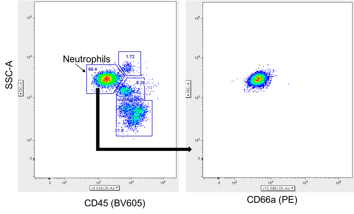
Application: Flow CytometrySample Tested: Peripheral blood neutrophilsSpecies: HumanVerified Customer | Posted 07/27/2020This antibody was used to stain human peripheral whole blood for analysis by flow cytometry. Neutrophils were identified from total live CD45+ leukocytes and expression of CD66a was measured in the PE channel. Consistent fluorescence is observed after fixation in 4% PFA. Good antibody for staining granulocytes

There are no reviews that match your criteria.
Protocols
Find general support by application which include: protocols, troubleshooting, illustrated assays, videos and webinars.
- 7-Amino Actinomycin D (7-AAD) Cell Viability Flow Cytometry Protocol
- Extracellular Membrane Flow Cytometry Protocol
- Flow Cytometry Protocol for Cell Surface Markers
- Flow Cytometry Protocol for Staining Membrane Associated Proteins
- Flow Cytometry Staining Protocols
- Flow Cytometry Troubleshooting Guide
- Intracellular Flow Cytometry Protocol Using Alcohol (Methanol)
- Intracellular Flow Cytometry Protocol Using Detergents
- Intracellular Nuclear Staining Flow Cytometry Protocol Using Detergents
- Intracellular Staining Flow Cytometry Protocol Using Alcohol Permeabilization
- Intracellular Staining Flow Cytometry Protocol Using Detergents to Permeabilize Cells
- Propidium Iodide Cell Viability Flow Cytometry Protocol
- Protocol for Liperfluo
- Protocol for the Characterization of Human Th22 Cells
- Protocol for the Characterization of Human Th9 Cells
- Protocol: Annexin V and PI Staining by Flow Cytometry
- Protocol: Annexin V and PI Staining for Apoptosis by Flow Cytometry
- Troubleshooting Guide: Fluorokine Flow Cytometry Kits
- View all Protocols, Troubleshooting, Illustrated assays and Webinars
Associated Pathways
