Human Cytokeratin 17 Antibody Summary
Accession # Q04695
Applications
Please Note: Optimal dilutions should be determined by each laboratory for each application. General Protocols are available in the Technical Information section on our website.
Scientific Data
 View Larger
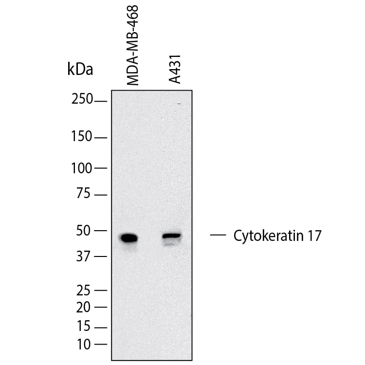
Detection of Human Cytokeratin 17 by Western Blot. Western Blot shows lysates of MDA‑MB‑468 human breast cancer cell line and A431 human epithelial carcinoma cell line. PVDF membrane was probed with 2 µg/ml of Mouse Anti-Human Cytokeratin 17 Monoclonal Antibody (Catalog # MAB11571) followed by HRP-conjugated Anti-Mouse IgG Secondary Antibody (Catalog # HAF018). A specific band was detected for Cytokeratin 17 at approximately 53 kDa (as indicated). This experiment was conducted under reducing conditions and using Western Blot Buffer Group 1.
 View Larger
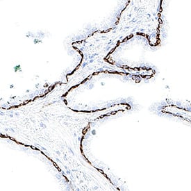
Detection of Cytokeratin 17 in Human Prostate. Cytokeratin 17 was detected in immersion fixed paraffin-embedded sections of human prostate using Mouse Anti-Human Cytokeratin 17 Monoclonal Antibody (Catalog # MAB11571) at 5 µg/ml for 1 hour at room temperature followed by incubation with the Anti-Mouse IgG VisUCyte™ HRP Polymer Antibody (Catalog # VC001) or the HRP-conjugated Anti-Mouse IgG Secondary Antibody (Catalog # HAF007). Before incubation with the primary antibody, tissue was subjected to heat-induced epitope retrieval using VisUCyte Antigen Retrieval Reagent-Basic (Catalog # VCTS021). Tissue was stained using DAB (brown) and counterstained with hematoxylin (blue). Specific staining was localized to the cytoplasm. View our protocol for Chromogenic IHC Staining of Paraffin-embedded Tissue Sections.
 View Larger
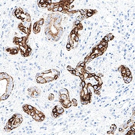
Detection of Cytokeratin 17 in Human Squamous Cell Carcinoma. Cytokeratin 17 was detected in immersion fixed paraffin-embedded sections of human squamous cell carcinoma using Mouse Anti-Human Cytokeratin 17 Monoclonal Antibody (Catalog # MAB11571) at 5 µg/ml for 1 hour at room temperature followed by incubation with the Anti-Mouse IgG VisUCyte™ HRP Polymer Antibody (Catalog # VC001) or the HRP-conjugated Anti-Mouse IgG Secondary Antibody (Catalog # HAF007). Before incubation with the primary antibody, tissue was subjected to heat-induced epitope retrieval using VisUCyte Antigen Retrieval Reagent-Basic (Catalog # VCTS021). Tissue was stained using DAB (brown) and counterstained with hematoxylin (blue). Specific staining was localized to the cytoplasm. View our protocol for Chromogenic IHC Staining of Paraffin-embedded Tissue Sections.
 View Larger
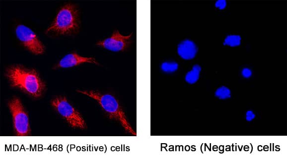
Detection of Cytokeratin 17 in MDA-MB-468 cells (Positive) and Ramos cells (Negative). Cytokeratin 17 was detected in fixed MDA‑MB‑468 human breast cancer cell line (Positive) and absent in Ramos human Burkitt's lymphoma cell line (Negative) using Mouse Anti-Human Cytokeratin 17 Monoclonal Antibody (Catalog # MAB11571) at 8 µg/ml for 3 hours at room temperature. Cells were stained using the NorthernLights™ 557-conjugated Anti-Mouse IgG Secondary Antibody (red; Catalog # NL007) and counterstained with DAPI (blue). Specific staining was localized to the cytoplasm. View our protocol for Fluorescent ICC Staining of Cells on Coverslips.
 View Larger
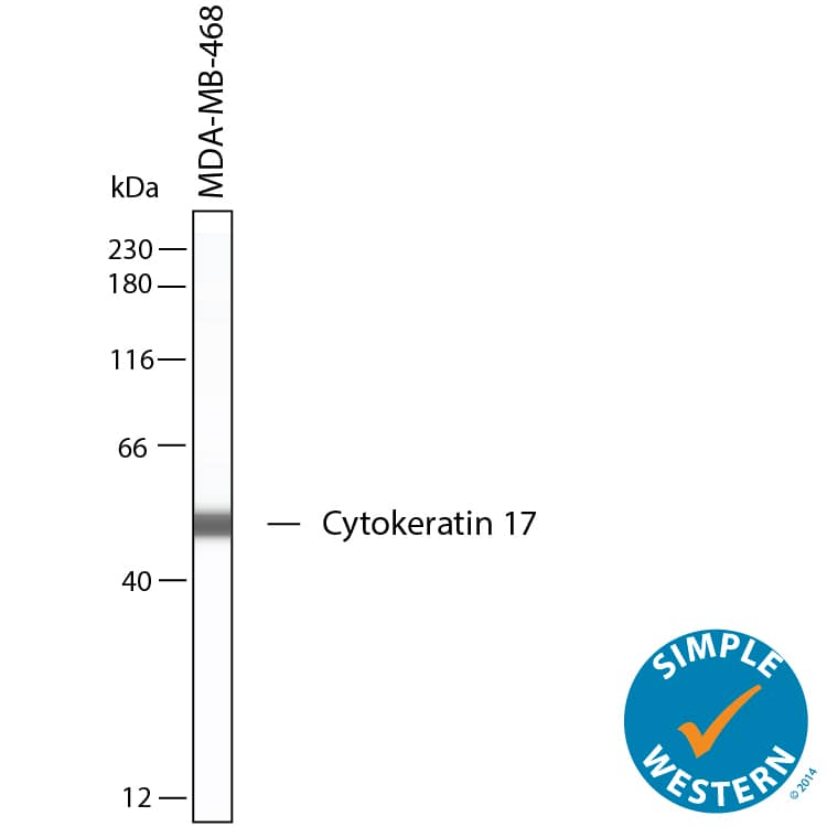
Detection of Human Cytokeratin 17 by Simple WesternTM. Simple Western shows lysates of MDA‑MB‑468 human breast cancer cell line, loaded at 0.5 mg/ml. A specific band was detected for Cytokeratin 17 at approximately 53 kDa (as indicated) using 100 µg/mL of Mouse Anti-Human Cytokeratin 17 Monoclonal Antibody (Catalog # MAB11571). This experiment was conducted under reducing conditions and using the 12‑230 kDa separation system.
Reconstitution Calculator
Preparation and Storage
- 12 months from date of receipt, -20 to -70 °C as supplied.
- 1 month, 2 to 8 °C under sterile conditions after reconstitution.
- 6 months, -20 to -70 °C under sterile conditions after reconstitution.
Background: Cytokeratin 17
Cytokeratin 17 (KRT17) is a 48 kDa, 432 aa type I keratin that has been studied in several types of cancer. KRT17 promotes epithelial cell proliferation and tumor growth in skin. Many studies have shown KRT17 overexpression in many cancers including cervical, oral, ovarian, gastric, lung and pancreatic cancer among others. In certain types of breast cancer, KRT17 overexpression has been associated with poor prognosis. KRT17 expression is closely associated with prognosis in cancer and can be a novel therapeutic target.
- Tang S, Liu W, Yong L, Liu D, Lin X, Huang Y, Wang H, Cai F. Reduced Expression of KRT17 Predicts Poor Prognosis in HER2high Breast Cancer. Biomolecules. 2022 Aug 25;12(9):1183. doi: 10.3390/biom12091183. PMID: 36139022; PMCID: PMC9496156.
- Hu H, Xu DH, Huang XX, Zhu CC, Xu J, Zhang ZZ, Zhao G. Keratin17 Promotes Tumor Growth and is Associated with Poor Prognosis in Gastric Cancer. J Cancer. 2018 Jan 1;9(2):346-357. doi: 10.7150/jca.19838. PMID: 29344281; PMCID: PMC5771342.
- Li D, Ni XF, Tang H, Zhang J, Zheng C, Lin J, Wang C, Sun L, Chen B. KRT17 Functions as a Tumor Promoter and Regulates Proliferation, Migration and Invasion in Pancreatic Cancer via mTOR/S6k1 Pathway. Cancer Manag Res. 2020 Mar 19;12:2087-2095. doi: 10.2147/CMAR.S243129. PMID: 32256116; PMCID: PMC7090205.
Product Datasheets
FAQs
No product specific FAQs exist for this product, however you may
View all Antibody FAQsReviews for Human Cytokeratin 17 Antibody
There are currently no reviews for this product. Be the first to review Human Cytokeratin 17 Antibody and earn rewards!
Have you used Human Cytokeratin 17 Antibody?
Submit a review and receive an Amazon gift card.
$25/€18/£15/$25CAN/¥75 Yuan/¥2500 Yen for a review with an image
$10/€7/£6/$10 CAD/¥70 Yuan/¥1110 Yen for a review without an image
