Human Drebrin 1 Antibody Summary
Asn482-Asp649 (Ser553Pro)
Accession # Q16643
Customers also Viewed
Applications
Please Note: Optimal dilutions should be determined by each laboratory for each application. General Protocols are available in the Technical Information section on our website.
Scientific Data
 View Larger
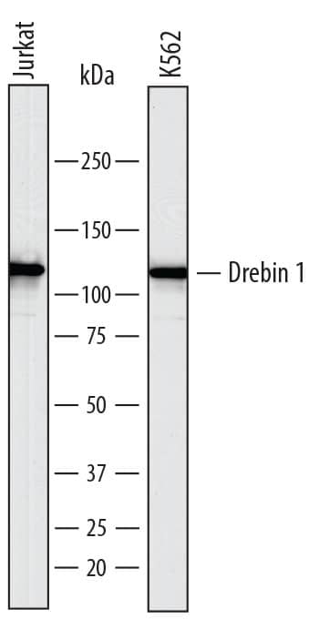
Detection of Human Drebrin 1 by Western Blot. Western blot shows lysates of Jurkat human acute T cell leukemia cell line and K562 human chronic myelogenous leukemia cell line. PVDF membrane was probed with 1 µg/mL of Sheep Anti-Human Drebrin 1 Antigen Affinity-purified Polyclonal Antibody (Catalog # AF7739) followed by HRP-conjugated Anti-Sheep IgG Secondary Antibody (Catalog # HAF016). A specific band was detected for Drebrin 1 at approximately 120 kDa (as indicated). This experiment was conducted under reducing conditions and using Immunoblot Buffer Group 1.
 View Larger
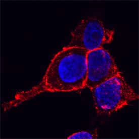
Drebrin 1 in HeLa Human Cell Line. Drebrin 1 was detected in immersion fixed HeLa human cervical epithelial carcinoma cell line using Sheep Anti-Human Drebrin 1 Antigen Affinity-purified Polyclonal Antibody (Catalog # AF7739) at 15 µg/mL for 3 hours at room temperature. Cells were stained using the NorthernLights™ 557-conjugated Anti-Sheep IgG Secondary Antibody (red; Catalog # NL010) and counterstained with DAPI (blue). Specific staining was localized to plasma membranes. View our protocol for Fluorescent ICC Staining of Cells on Coverslips.
Preparation and Storage
- 12 months from date of receipt, -20 to -70 °C as supplied.
- 1 month, 2 to 8 °C under sterile conditions after reconstitution.
- 6 months, -20 to -70 °C under sterile conditions after reconstitution.
Background: Drebrin 1
Drebrin 1 (DBN-1 [developmentally-regulated brain protein1]; also drebrin-E/E2 [Embryonic]) is an intracellular member of the ADF-H (actin-depolymerizing factor-H) family of actin binding proteins. Although its predicted MW is 72 kDa, it runs anomalously at 115-116 kDa in SDS-PAGE. It is expressed by neurons, gastric Parietal cells, astrocytes, distal convoluted tubule epithelium and proton-secreting intercalated cells of the renal collecting duct. Drebrin 1 interacts with multiple partners near the membrane. It links connexin-43 and F-actin, thereby stabilizing membrane gap junctions. It also binds to EB3 (end-binding protein 3) on microtubules, facilitating actin-microtubule interactions. Human Drebrin 1 is 649 amino acids (aa) in length. It contains one actin depolymerizing homology domain (aa 3-134), an actin-binding region (≈ aa 150-300), and two HOMER binding motifs (aa 539-543 and 617-621). There are at least 10 utilized Ser/Thr phosphorylation sites and one utilized Tyr phosphorylation site. Alternative splicing generates drebrin-A (Adult), a 124-126 kDa isoform that contains a 46 aa insert after Gly319. Drebrin-A is found in neurons and possibly podocytes, and is associated with dendritic spines where it inhibits the interaction of F-actin with alpha -actinin and tropomyosin. This favors the generation of excitatory impulses in neurons. Three other potential isoform variants are noted. One utilizes an alternative start site at Met64, a second shows a 60 aa substitution for aa 1-110, and a third contains a 28 aa substitution for aa 4-29. Over aa 482-649, human Drebrin 1 shares 84% aa sequence identity with mouse Drebrin 1.
Product Datasheets
FAQs
No product specific FAQs exist for this product, however you may
View all Antibody FAQsReviews for Human Drebrin 1 Antibody
There are currently no reviews for this product. Be the first to review Human Drebrin 1 Antibody and earn rewards!
Have you used Human Drebrin 1 Antibody?
Submit a review and receive an Amazon gift card.
$25/€18/£15/$25CAN/¥75 Yuan/¥2500 Yen for a review with an image
$10/€7/£6/$10 CAD/¥70 Yuan/¥1110 Yen for a review without an image

