Human EMP3 Antibody Summary
Met1-Glu163
Accession # P54852
Applications
Please Note: Optimal dilutions should be determined by each laboratory for each application. General Protocols are available in the Technical Information section on our website.
Scientific Data
 View Larger
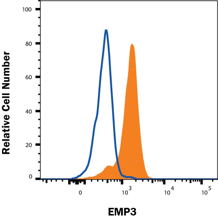
Detection of EMP3 in Human PBMCs by Flow Cytometry. Human peripheral blood mononuclear cells were stained with Mouse Anti-Human EMP3 Monoclonal Antibody (Catalog # MAB4505, filled histogram) or isotype control antibody (MAB0041, open histogram), followed by Phycoerythrin-conjugated Anti-Mouse IgG F(ab')2Secondary Antibody (F0102B). To facilitate intracellular staining, cells were fixed with paraformaldehyde and permeabilized with saponin.
 View Larger
Detection of EMP3 in PBMC monocytes by Flow Cytometry. PBMC monocytes were stained with Mouse Anti-Human EMP3 Monoclonal Antibody (Catalog # MAB4505, filled histogram) or isotype control antibody (Catalog # MAB0041, open histogram), followed by Phycoerythrin-conjugated Anti-Mouse IgG Secondary Antibody (Catalog # F0102B). To facilitate intracellular staining, cells were fixed and permeabilized with Flow Cytometry Fixation Buffer (Catalog # FC004). View our protocol for Staining Intracellular Molecules.
Reconstitution Calculator
Preparation and Storage
- 12 months from date of receipt, -20 to -70 °C as supplied.
- 1 month, 2 to 8 °C under sterile conditions after reconstitution.
- 6 months, -20 to -70 °C under sterile conditions after reconstitution.
Background: EMP3
Epithelial membrane protein 3 (EMP3) is a multipass transmembrane protein in the peripheral myelin protein 22 family. It is expressed as a 20‑25 kDa molecule depending on the degree of glycosylation. EMP3 interacts with the P2X7 purinergic receptor on monocytes. Its dysregulation is associated with glioma and neuroblastoma. Human EMP3 shares 93% aa sequence identity with mouse and rat EMP3.
Product Datasheets
Citation for Human EMP3 Antibody
R&D Systems personnel manually curate a database that contains references using R&D Systems products. The data collected includes not only links to publications in PubMed, but also provides information about sample types, species, and experimental conditions.
1 Citation: Showing 1 - 1
-
Disruption of the tumour-associated EMP3 enhances erythroid proliferation and causes the MAM-negative phenotype
Authors: N Thornton, V Karamatic, L Tilley, CA Green, CL Tay, RE Griffiths, BK Singleton, F Spring, P Walser, AG Alattar, B Jones, R Laundy, JR Storry, M Möller, L Wall, R Charlewood, CM Westhoff, C Lomas-Fran, V Yahalom, U Feick, A Seltsam, B Mayer, ML Olsson, DJ Anstee
Nat Commun, 2020-07-16;11(1):3569.
Species: Human
Sample Types: Whole Cells
Applications: Flow Cytometry, Neutralization
FAQs
-
Why do you recommend intracellular staining with a flow antibody to a membrane expressed protein?
The antibody may recognize an intracellular portion of the protein,or an abundance of the protein may be in an intracellular location. In our hands, the intracellular staining worked better than surface staining.
Reviews for Human EMP3 Antibody
There are currently no reviews for this product. Be the first to review Human EMP3 Antibody and earn rewards!
Have you used Human EMP3 Antibody?
Submit a review and receive an Amazon gift card.
$25/€18/£15/$25CAN/¥75 Yuan/¥2500 Yen for a review with an image
$10/€7/£6/$10 CAD/¥70 Yuan/¥1110 Yen for a review without an image

