Human GATA-1 PE-conjugated Antibody

Catalog # Availability Size / Price Qty
IC1779P
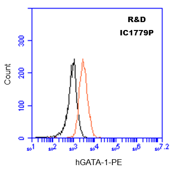
Detection of GATA‑1 in MG‑63 Human Cell Line by Flow Cytometry.
1 Image
Product Details
FAQs
Supplemental Products
Reviews (1)

Human GATA-1 PE-conjugated Antibody Summary

Species Reactivity
Human
Specificity
Detects human GATA-1 in direct ELISAs. In Western blots, this antibody does not cross-react with recombinant human (rh) GATA-2 or rhGATA-6.
Source
Monoclonal Rat IgG2B Clone # 234732
Purification
Protein A or G purified from hybridoma culture supernatant
Immunogen
E. coli-derived recombinant human GATA-1
Met1-Ser413
Accession # P15976
Formulation
Supplied in a saline solution containing BSA and Sodium Azide.
Label
Phycoerythrin (Excitation= 488 nm, Emission= 565-605 nm)
Purity
Protein A or G purified from hybridoma culture supernatant

Applications

Recommended Concentration
Sample
Intracellular Staining by Flow Cytometry
10 µL/106 cells
See below

Please Note: Optimal dilutions should be determined by each laboratory for each application. General Protocols are available in the Technical Information section on our website.

Scientific Data

Intracellular Staining by Flow Cytometry Detection of GATA-1 antibody in MG-63 Human Cell Line antibody by Flow Cytometry. View Larger

Detection of GATA‑1 in MG‑63 Human Cell Line by Flow Cytometry. MG-63 human osteosarcoma cell line was stained with Rat Anti-Human GATA-1 PE-conjugated Monoclonal Antibody (Catalog # IC1779P, filled histogram) or isotype control antibody (Catalog # IC013P, open histogram). To facilitate intracellular staining, cells were fixed with Flow Cytometry Fixation Buffer (Catalog # FC004) and permeabilized with Flow Cytometry Permeabilization/Wash Buffer I (Catalog # FC005). View our protocol for Staining Intracellular Molecules.

Reconstitution Calculator

Reconstitution Calculator

The reconstitution calculator allows you to quickly calculate the volume of a reagent to reconstitute your vial. Simply enter the mass of reagent and the target concentration and the calculator will determine the rest.

=
÷

Preparation and Storage

Shipping
The product is shipped with polar packs. Upon receipt, store it immediately at the temperature recommended below.
Stability & Storage
Store the unopened product at 2 - 8° C. Do not use past expiration date. Protect from light.

Background: GATA-1

GATA-1 is the founding member of the GATA family of transcription factors, which bind to the consensus DNA sequence (A/T) GATA (A/G) to control diverse tissue‑specific programs of gene expression and morphogenesis. GATA-1 is expressed in blood forming cells. It interacts with several additional proteins to activate or repress gene expression and is essential for erythropoiesis (1).

References
  1. Patient, R.K. and J. McGhee (2002) Curr. Opin. Genet. Dev. 12:416.
Entrez Gene IDs
2623 (Human); 14460 (Mouse)
Alternate Names
Eryf1; ERYF1GATA-binding protein 1 (globin transcription factor 1); GATA binding protein 1 (globin transcription factor 1); GATA1; GATA-1; GATA-1erythroid transcription factor; GATA-binding factor 1; GF-1; GF1globin transcription factor 1; NF-E1 DNA-binding protein; NFE1erythroid transcription factor 1; transcription factor GATA1; XLTT

Product Datasheets

You must select a language.

x

FAQs

No product specific FAQs exist for this product, however you may

View all Antibody FAQs
Loading...

Reviews for Human GATA-1 PE-conjugated Antibody

Average Rating: 5 (Based on 1 Review)

5 Star
100%
4 Star
0%
3 Star
0%
2 Star
0%
1 Star
0%

Have you used Human GATA-1 PE-conjugated Antibody?

Submit a review and receive an Amazon gift card.

$25/€18/£15/$25CAN/¥75 Yuan/¥2500 Yen for a review with an image

$10/€7/£6/$10 CAD/¥70 Yuan/¥1110 Yen for a review without an image

Submit a Review

Filter by:


Human GATA-1 PE-conjugated Antibody
By Anonymous on 03/27/2017
Application: Flow Sample Tested: MCF-7 human breast cancer cell line Species: Human

Intracellular detection of GATA-1 in MCF-7 cells using the PE-conjugated Human GATA-1 antibody (#IC1779P, orange) at 10 uL/10^6 cells, or Isotype Control antibody (black). Cells were fixed with 2% PFA for 10 min and permeabilized with 0.2% Tween-20 for 20 min prior to staining. Staining buffer: 0.2% Tween-20.