Human GATA-3 Antibody Summary
Pro135-Ser258
Accession # P23771
Applications
Please Note: Optimal dilutions should be determined by each laboratory for each application. General Protocols are available in the Technical Information section on our website.
Scientific Data
 View Larger
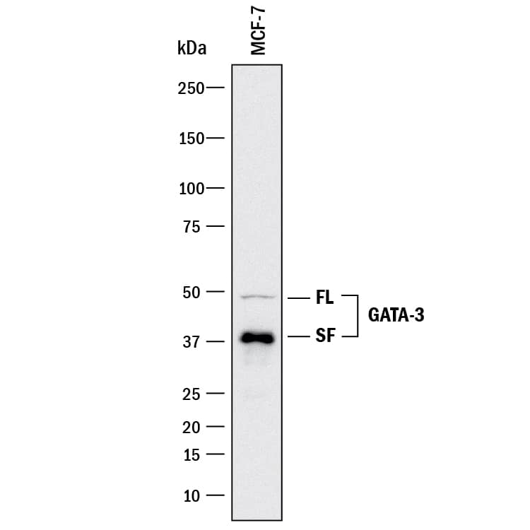
Detection of Human GATA‑3 by Western Blot. Western blot shows lysates of MCF-7 human breast cancer cell line. PVDF membrane was probed with 2 µg/mL of Mouse Anti-Human GATA-3 Monoclonal Antibody (Catalog # MAB26052) followed by HRP-conjugated Anti-Mouse IgG Secondary Antibody (HAF018). Specific bands were detected for GATA-3 full length (FL) at approximately 50 kDa and the splice form (SF) at approximately 37 kDa (as indicated). This experiment was conducted under reducing conditions and using Immunoblot Buffer Group 3.
 View Larger
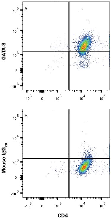
Detection of GATA‑3 in Human PBMCs by Flow Cytometry. Human peripheral blood mononuclear cells (PBMCs) stimulated to induce Th2 cells were stained with Mouse Anti-Human CD4 PE-conjugated Monoclonal Antibody (FAB3791P) and either (A) Mouse Anti-Human GATA-3 Monoclonal Antibody (Catalog # MAB26052) or (B) Mouse IgG2BFlow Cytometry Isotype Control (MAB0041) followed by Allophycocyanin-conjugated Anti-Mouse IgG Secondary Antibody (F0101B). To facilitate intracellular staining, cells were fixed and permeabilized with FlowX FoxP3 Fixation & Permeabilization Buffer Kit (FC012). View our protocol for Staining Intracellular Molecules.
 View Larger
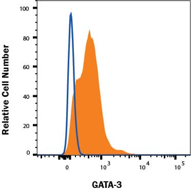
Detection of GATA‑3 in MCF-7 cells by Flow Cytometry. MCF-7 cells were stained with Mouse Anti-Human GATA‑3 Monoclonal Antibody (Catalog # MAB26052, filled histogram) or isotype control antibody (Catalog # MAB004, open histogram), followed by Allophycocyanin-conjugated Anti-Mouse IgG Secondary Antibody (Catalog # F0101B). To facilitate intracellular staining, cells were fixed with FC012 and permeabilized with FoxP3 Perm. View our protocol for Staining Intracellular Molecules.
Reconstitution Calculator
Preparation and Storage
- 12 months from date of receipt, -20 to -70 °C as supplied.
- 1 month, 2 to 8 °C under sterile conditions after reconstitution.
- 6 months, -20 to -70 °C under sterile conditions after reconstitution.
Background: GATA-3
GATA-3 belongs to the GATA family of transcription factors, which bind to the consensus DNA sequence (A/T) GATA (A/G) to control diverse tissue-specific programs of gene expression and morphogenesis. It is widely expressed in mesodermal- and endodermal-derived tissues. GATA-3 has been shown to be an essential regulator for immune cell function, sympathetic neuron development and the maintenance of the differentiated state in epithelial cells.
Product Datasheets
FAQs
No product specific FAQs exist for this product, however you may
View all Antibody FAQsReviews for Human GATA-3 Antibody
There are currently no reviews for this product. Be the first to review Human GATA-3 Antibody and earn rewards!
Have you used Human GATA-3 Antibody?
Submit a review and receive an Amazon gift card.
$25/€18/£15/$25CAN/¥75 Yuan/¥2500 Yen for a review with an image
$10/€7/£6/$10 CAD/¥70 Yuan/¥1110 Yen for a review without an image


