Human Granulysin Antibody Summary
Arg23-Leu145
Accession # P22749
*Small pack size (-SP) is supplied either lyophilized or as a 0.2 µm filtered solution in PBS.
Applications
Please Note: Optimal dilutions should be determined by each laboratory for each application. General Protocols are available in the Technical Information section on our website.
Scientific Data
 View Larger
Detection of Granulysin in Human PBMCs by Flow Cytometry. Human peripheral blood mononuclear cells were treated for 24 hr with 50 ng/ml PMA and 200 ng/ml Ca2+ionomycin then stained with Goat Anti-Human Granulysin Antigen Affinity-purified Polyclonal Antibody (Catalog # AF3138, filled histogram) or control antibody (AB-108-C, open histogram), followed by Phycoerythrin-conjugated Anti-Goat IgG Secondary Antibody (F0107).
 View Larger
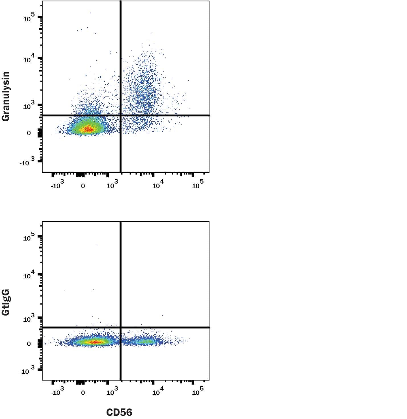
Detection of Granulysin in human PBMCs by Flow Cytometry Human PBMCs were stained with Rabbit Anti-Human NCAM‑1/CD56 PE‑conjugated Monoclonal Antibody (Catalog # FAB24086P) and either (A) Goat Anti-Human Granulysin Antigen Affinity-purified Polyclonal Antibody (Catalog # AF3138) or (B) isotype control antibody (Catalog # AB-108-C) followed by Allophycocyanin-conjugated Anti-Goat IgG Secondary Antibody (Catalog # F0108). To facilitate intracellular staining, cells were fixed with Flow Cytometry Fixation Buffer (Catalog # FC004) and permeabilized with Flow Cytometry Permeabilization/Wash Buffer I (Catalog # FC005). View our protocol for Staining Intracellular Molecules.
Reconstitution Calculator
Preparation and Storage
- 12 months from date of receipt, -20 to -70 °C as supplied.
- 1 month, 2 to 8 °C under sterile conditions after reconstitution.
- 6 months, -20 to -70 °C under sterile conditions after reconstitution.
Background: Granulysin
Granulysin (formerly NKG5 or Lymphokine LAG-2) is a member of the saposin-like protein (SAPLIP) family of membrane disrupting proteins (1). Granulysin is expressed in granules of natural killer and activated cytotoxic T cells. It exhibits cytolytic activity against intracellular or extracellular microbes and also tumors, either alone or in synergy with perforin (2). Human granulysin has structural similarity and 30 - 40% aa identity to granulysins and NK-lysins of other mammals such as bovine, porcine and canine; similar peptides in rodents have not been identified (1). The 15 kDa unglycosylated protein contains five helical domains; helix 2 and 3 contain 9 arginines and one cysteine critical for activity. Peptides of either helix 2 or 3 will lyse bacteria, while helix 3 is needed to lyse tumor targets (3, 4). One isoform designated 519 uses a different start codon, has no signal peptide sequence and is poorly expressed (5). A post-translationally processed 9 kDa form is present in acidified granules and is less lytic than the 15 kDa form at granule pH (6). IL-15 is necessary and sufficient for granulysin upregulation in CD8 T cells (2). Nanomolar granulysin promotes chemotaxis and increases production of chemokines by monocytic cells, while micromolar local concentrations are needed for lysis (7). Experimental evidence supports the following mechanism for activity against intracellular pathogens (8). First, granulysin forms clusters in lipid rafts due to interaction of positive charges in helices 2-3 with acidic sphingolipids. After endocytosis, early endosomes fuse with phagosomes, probably regulated by small GTPase rab5. Granulysin binds microbial membranes through charge interactions and disrupts them, probably via scissoring actions of granulysin molecules (9, 10).
- Clayberger, C. and A.M. Krensky (2003) Curr. Opin. Immunol. 15:560.
- Ma, L.L. et al. (2002) J. Immunol. 169:5787.
- Wang, Z. et al. (2000) J. Immunol. 165:1486.
- Linde, C.M.A. et al. (2005) Infect. Immun. 73:6332.
- Yabe, T. et al. (1990) J. Exp. Med. 172:1159.
- Hanson, D.A. et al. (1999) Mol. Immunol. 36:413.
- Deng, A. et al. (2005) J. Immunol. 174:5243.
- Walch, M. et al. (2005) J. Immunol. 174:4220.
- Ernst, W.A. et al. (2000) J. Immunol. 165:7102.
- Anderson, D.H. et al. (2003) J. Mol. Biol. 325:355.
Product Datasheets
Citations for Human Granulysin Antibody
R&D Systems personnel manually curate a database that contains references using R&D Systems products. The data collected includes not only links to publications in PubMed, but also provides information about sample types, species, and experimental conditions.
2
Citations: Showing 1 - 2
Filter your results:
Filter by:
-
Analysis of Cytotoxic Granules and Constitutively Produced Extracellular Vesicles from Large Granular Lymphocytic Leukemia Cell Lines
Authors: Ploeger, L;Kaleja, P;Tholey, A;Lettau, M;Janssen, O;
Cells
Species: Human
Sample Types: Cell Lysates
Applications: Immunoprecipitation -
An E. coli display method for characterization of peptide-sensor kinase interactions
Authors: KR Brink, MG Hunt, AM Mu, K Groszman, KV Hoang, KP Lorch, BH Pogostin, JS Gunn, JJ Tabor
Nature Chemical Biology, 2022-12-08;0(0):.
Species: Human
Sample Types: Cell Lysates
FAQs
No product specific FAQs exist for this product, however you may
View all Antibody FAQsReviews for Human Granulysin Antibody
There are currently no reviews for this product. Be the first to review Human Granulysin Antibody and earn rewards!
Have you used Human Granulysin Antibody?
Submit a review and receive an Amazon gift card.
$25/€18/£15/$25CAN/¥75 Yuan/¥2500 Yen for a review with an image
$10/€7/£6/$10 CAD/¥70 Yuan/¥1110 Yen for a review without an image

