Human JAM-C Antibody Summary
Val32-Asn241 (Ala149Pro)
Accession # Q9BX67
Applications
Please Note: Optimal dilutions should be determined by each laboratory for each application. General Protocols are available in the Technical Information section on our website.
Scientific Data
 View Larger
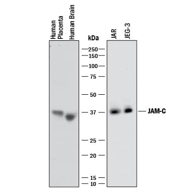
Detection of Human JAM‑C by Western Blot. Western blot shows lysates of human placenta tissue, human brain tissue, JAR human choriocarcinoma cell line, and JEG-3 human epithelial choriocarcinoma cell line. PVDF membrane was probed with 2 µg/mL of Mouse Anti-Human JAM-C Monoclonal Antibody (Catalog # MAB1189) followed by HRP-conjugated Anti-Mouse IgG Secondary Antibody (Catalog # HAF018). A specific band was detected for JAM-C at approximately 36-38 kDa (as indicated). This experiment was conducted under reducing conditions and using Immunoblot Buffer Group 1.
 View Larger
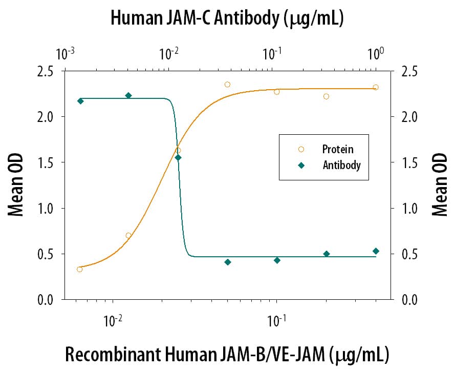
Cell Adhesion Mediated by JAM‑C and Neutralization by Human JAM‑C Antibody. Recombinant Human JAM-B/VE-JAM Fc Chimera, immobilized onto a microplate previously coated with Goat Anti-Human IgG Fc (Catalog # G-102-C), supports the adhesion of the J45.01 human acute lymphoblastic leukemia T lymphocyte cell line in a dose-dependent manner (orange line), as measured by endogenous cellular lysosomal acid phosphatase activity. Adhesion elicited by Recombinant Human JAM-B/VE-JAM Fc Chimera (0.2 µg/mL) is neutralized (green line) by increasing concentrations of Mouse Anti-Human JAM-C Monoclonal Antibody (Catalog # MAB1189). The ND50 is typically 0.01-0.05 µg/mL.
Reconstitution Calculator
Preparation and Storage
- 12 months from date of receipt, -20 to -70 °C as supplied.
- 1 month, 2 to 8 °C under sterile conditions after reconstitution.
- 6 months, -20 to -70 °C under sterile conditions after reconstitution.
Background: JAM-C
The family of juctional adhesion molecules (JAM), comprising at least three members, are type I transmembrane receptors belonging to the immunoglobulin (Ig) superfamily (1, 2). These proteins are localized in the tight junctions between endothelial cells or epithelial cells. Some family members are also found on blood leukocytes and platelets. Human JAM-C cDNA predicts a 310 amino acid (aa) residue precursor protein with a putative 31 aa signal peptide, a 210 aa extracellular region containing two Ig domains, a 23 aa transmembrane domain and a 46 aa cytoplasmic domain containing a PDZ-binding motif and a PKC phosphorylation site (3, 4). Human JAM-C shares 86% aa sequence identity with its mouse homologue. It also shares approximately 36% and 32% aa sequence homology with human JAM-B and JAM-A, respectively (3‑5). Human JAM-C shows widespread tissue expression and the highest levels are found in the placenta, brain, kidney and heart. JAM-C is expressed on endothelial cells of high endothelial venules in human tonsil. It is also expressed on platelets, T-cells and NK cells (3‑5). Unlike other JAM family members, JAM-C forms only weak homotypic interactions. JAM-C binds to JAM-B to facilitate the interactions between JAM-B and the integrin alpha4beta1 (6). This heterotypic interaction between leukocyte JAM-C and endothelial JAM-B may play a role in regulating leukocyte transmigration (5). On platelets, JAM-C is a counter-receptor for the leukocyte integrin Mac-1(CD11b/CD18) (7). JAM-C has also been identified as a strong candidate gene for hypoplastic left heart syndrome (8).
The nomenclature used for the JAM family proteins is confusing. VE-JAM has been referred to in the literature variously as JAM-B or JAM-C. Until further clarification, R&D Systems has adopted the nomenclature where both mouse and human VE-JAM are referred to as JAM-B. Under this system, JAM-C refers to the protein encoded by the gene localized to human chromosome 11.
- Chavakis, T. et al. (2003) Thromb. Haemost. 89:13.
- Aurand-Lions, M. et al. (2001) Blood 98:3699.
- Arrate, M.P. et al. (2001) J. Biol. Chem. 276:45826.
- Liang, T. et al. (2002) J. Immunol. 168:1618.
- Johnson-Leger, C. et al. (2002) Blood 100:25793.
- Cunningham, A. et al. (2002) J Biol. Chem. 277:27589.
- Santoso, S. et al. (2002) J. Exp. Med. 196:679.
- Phillips, H.M. et al. (2002) Genomics 79:475.
Product Datasheets
Citations for Human JAM-C Antibody
R&D Systems personnel manually curate a database that contains references using R&D Systems products. The data collected includes not only links to publications in PubMed, but also provides information about sample types, species, and experimental conditions.
3
Citations: Showing 1 - 3
Filter your results:
Filter by:
-
Constitutive and functionally relevant expression of JAM-C on platelets.
Authors: Erpenbeck L, Rubant S, Hardt K, Santoso S, Boehncke WH, Schon MP, Ludwig RJ
Thromb. Haemost., 2010-02-02;103(4):857-9.
Species: Human
Sample Types: Cell Lysates, Whole Cells
Applications: Neutralization, Western Blot -
Possible involvement of gap junctions in the barrier function of tight junctions of brain and lung endothelial cells.
Authors: Nagasawa K, Chiba H, Fujita H, Kojima T, Saito T, Endo T, Sawada N
J. Cell. Physiol., 2006-07-01;208(1):123-32.
Species: Porcine
Sample Types: Cell Lysates, Whole Cells
Applications: ICC, Western Blot -
Antibody blockade of junctional adhesion molecule-A in rabbit corneal endothelial tight junctions produces corneal swelling.
Authors: Mandell KJ, Holley GP, Parkos CA, Edelhauser HF
Invest. Ophthalmol. Vis. Sci., 2006-06-01;47(6):2408-16.
Species: Rabbit
Sample Types: Whole Tissue
Applications: IHC-Fr
FAQs
No product specific FAQs exist for this product, however you may
View all Antibody FAQsReviews for Human JAM-C Antibody
There are currently no reviews for this product. Be the first to review Human JAM-C Antibody and earn rewards!
Have you used Human JAM-C Antibody?
Submit a review and receive an Amazon gift card.
$25/€18/£15/$25CAN/¥75 Yuan/¥2500 Yen for a review with an image
$10/€7/£6/$10 CAD/¥70 Yuan/¥1110 Yen for a review without an image


