Human Lgr5/GPR49 Antibody Summary
Met1-Ile560
Accession # O75473
Applications
Please Note: Optimal dilutions should be determined by each laboratory for each application. General Protocols are available in the Technical Information section on our website.
Scientific Data
 View Larger
Detection of Lgr5/GPR49 in NS0 Mouse Cell Line Transfected with Human Lgr5/GPR49 and eGFP by Flow Cytometry. NS0 mouse myeloma cell line transfected with human Lgr5/GPR49 and eGFP was stained with and either (A) Mouse Anti-Human Lgr5/GPR49 Monoclonal Antibody (Catalog # MAB8078) or (B) Mouse IgG2AIsotype Control (Catalog # MAB003) followed by Allophycocyanin-conjugated Anti-Mouse IgG Secondary Antibody (Catalog # F0101B).
 View Larger
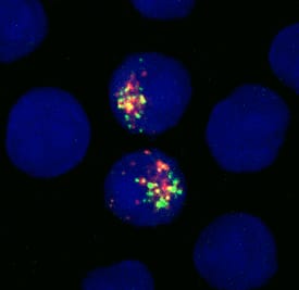
Lgr5/GPR49 in NSO Mouse Cell Line Transfected with Human Lgr5/GPR49. Lgr5/GPR49 was detected in immersion fixed NSO mouse myeloma cell line transfected with GFP (green) tagged human LGR5 using Mouse Anti-Human Lgr5/GPR49 Monoclonal Antibody (Catalog # MAB8078) at 10 µg/mL for 3 hours at room temperature. Cells were stained using the NorthernLights™ 557-conjugated Anti-Mouse IgG Secondary Antibody (red; Catalog # NL007) and counterstained with DAPI (blue). Specific staining was localized to cytoplasm. View our protocol for Fluorescent ICC Staining of Non-adherent Cells.
 View Larger
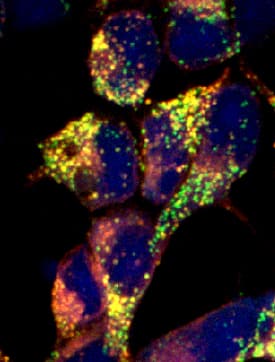
Lgr5/GPR49 in HEK293 Human Cell Line Transfected with Mouse Lgr5/GPR49. Lgr5/GPR49 was detected in immersion fixed HEK293 human embryonic kidney cell line transfected with GFP (green) tagged mouse LGR5 using Mouse Anti-Human Lgr5/GPR49 Monoclonal Antibody (Catalog # MAB8078) at 10 µg/mL for 3 hours at room temperature. Cells were stained using the NorthernLights™ 557-conjugated Anti-Mouse IgG Secondary Antibody (red; Catalog # NL007) and counterstained with DAPI (blue). Specific staining was localized to cell surfaces and cytoplasm. View our protocol for Fluorescent ICC Staining of Cells on Coverslips.
 View Larger
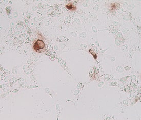
Lgr5/GPR49 in Human Cells Transfected with Human Lgr5/GPR49. Lgr5/GPR49 was detected in MYC tagged human transfectants using Mouse Anti-Human Lgr5/GPR49 Monoclonal Antibody (Catalog # MAB8078). Cells were stained using an anti-mouse HRP-conjugated secondary antibody (brown). Specific staining was localized to cytoplasm.Image courtesy of Dr. Hans Clevers, Hubrecht Institute, The Netherlands.
 View Larger
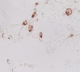
Lgr5/GPR49 in Mouse Cells Transfected with Mouse Lgr5/GPR49. Lgr5/GPR49 was detected in immersion fixed MYC tagged mouse transfectants using Mouse Anti-Human Lgr5/GPR49 Monoclonal Antibody (Catalog # MAB8078). Cells were stained using an anti-mouse HRP-conjugated secondary antibody (brown). Specific staining was localized to cytoplasm.Image courtesy of Dr. Hans Clevers, Hubrecht Institute, The Netherlands.
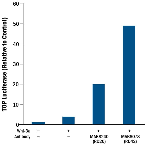 View Larger
Human Lgr5/GPR49 Antibody Induces Activity. Mouse Anti-Human Lgr5/GPR49 Monoclonal Antibody (Catalog # MAB8078) induces TOPflash activity in the HEK293 human embryonic kidney cell line stably expressing LGR5 in the presence of Wnt-3a, but in the absence of R-Spondins. (Data courtesy of Dr. Wim de Lau and Dr. Hans Clevers, Hubrecht Institute, The Netherlands. See Reference 1.)
Reconstitution Calculator
Preparation and Storage
- 12 months from date of receipt, -20 to -70 °C as supplied.
- 1 month, 2 to 8 °C under sterile conditions after reconstitution.
- 6 months, -20 to -70 °C under sterile conditions after reconstitution.
Background: Lgr5/GPR49
Leucine‑rich repeat G‑protein‑coupled receptor 5 (Lgr5), also called GPR49, is a 907 amino acid (aa), approximately 97 kDa (calculated), seven‑transmembrane glycoprotein receptor in the Lgr family of cell surface receptors. The subfamily of Lgrs comprising Lgr4, Lgr5, and Lgr6 are G‑protein‑independent mediators of the potentiating effect of R‑Spondins on Wnt signaling (2). Lgr5 binds and forms complexes with R‑Spondins, Frizzled Wnt receptors and LRP Wnt co‑receptors. The region of the human Lgr5 long extracellular domain used as an immunogen shares 90% amino acid sequence identity with mouse and rat Lgr5, respectively. Lgr5 is found on embryonic and adult epithelial stem cells (3). Lgr5+ stem cells can produce all epithelial cell types of the intestinal crypts (4). Abnormal LGR5 expression and regulation in stem cells might give rise to cancers such as intestinal, hepatocellular, pancreatic and ovarian carcinomas (5,6). This antibody has been referred to as "RD42" in Peng et al. (1).
- Peng et al. (2013) Cell Rep. 3(6):1885.
- de Lau et al. (2014) Genes Dev. 28(4):305.
- Barker et al. (2013) Development 140(12):2484.
- Clevers (2013) Nature 495(7439):53.
- Wu et al. (2014) Nat Commun 5:3149.
- Jang et al. (2013) PLoS One 8(12): e82390495.
Product Datasheets
Citations for Human Lgr5/GPR49 Antibody
R&D Systems personnel manually curate a database that contains references using R&D Systems products. The data collected includes not only links to publications in PubMed, but also provides information about sample types, species, and experimental conditions.
3
Citations: Showing 1 - 3
Filter your results:
Filter by:
-
3D bioprinted colorectal cancer models based on hyaluronic acid and signalling glycans
Authors: F Cadamuro, L Marongiu, M Marino, N Tamini, L Nespoli, N Zucchini, A Terzi, D Altamura, Z Gao, C Giannini, G Bindi, A Smith, F Magni, S Bertini, F Granucci, F Nicotra, L Russo
Carbohydrate polymers, 2022-11-30;302(0):120395.
Species: Human
Sample Types: Organoid
Applications: IHC -
Activin A-mediated epithelial de-differentiation contributes to injury repair in an in vitro gastrointestinal reflux model
Authors: C Roudebush, A Catala-Val, T Andl, GF Le Bras, CD Andl
Cytokine, 2019-07-29;123(0):154782.
Species: Human
Sample Types: Whole Tissue
Applications: IHC-P -
Musashi-1 promotes a cancer stem cell lineage and chemoresistance in colorectal cancer cells
Authors: GY Chiou, TW Yang, CC Huang, CY Tang, JY Yen, MC Tsai, HY Chen, N Fadhilah, CC Lin, YJ Jong
Sci Rep, 2017-05-19;7(1):2172.
Species: Human
Sample Types: Cell Lysates
Applications: Western Blot
FAQs
No product specific FAQs exist for this product, however you may
View all Antibody FAQsReviews for Human Lgr5/GPR49 Antibody
There are currently no reviews for this product. Be the first to review Human Lgr5/GPR49 Antibody and earn rewards!
Have you used Human Lgr5/GPR49 Antibody?
Submit a review and receive an Amazon gift card.
$25/€18/£15/$25CAN/¥75 Yuan/¥2500 Yen for a review with an image
$10/€7/£6/$10 CAD/¥70 Yuan/¥1110 Yen for a review without an image


