Human Matrilin-2 Antibody Summary
Arg24-Arg937
Accession # AAH10444
Applications
Please Note: Optimal dilutions should be determined by each laboratory for each application. General Protocols are available in the Technical Information section on our website.
Scientific Data
 View Larger
Matrilin‑2 in U2OS Human Cell Line. Matrilin‑2 was detected in immersion fixed U2OS human osteosarcoma cell line using Goat Anti-Human Matrilin‑2 Antigen Affinity-purified Polyclonal Antibody (Catalog # AF3044) at 5 µg/mL for 3 hours at room temperature. Cells were stained using the NorthernLights™ 557-conjugated Anti-Goat IgG Secondary Antibody (red; NL001) and counterstained with DAPI (blue). Specific staining was localized to cytoplasm. Staining was performed using our protocol for Fluorescent ICC Staining of Non-adherent Cells.
 View Larger
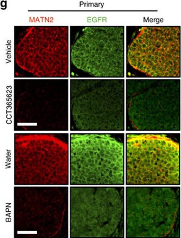
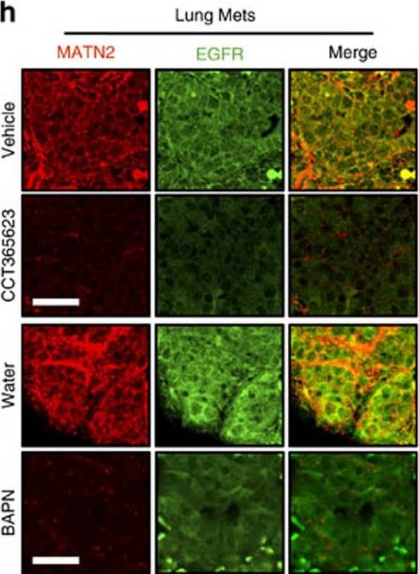
Detection of Mouse Matrilin-2 by Immunohistochemistry Chemical inhibition of LOX blocks tumour growth and metastasis.(a) Tumour growth in vehicle or CCT365623-treated MMTV-PyMT mice. Data are represented as mean±s.d. from six mice per group. **P<0.01, Student's t-test. (b) H&E stained lungs from vehicle or CCT365623 treated MMTV-PyMT mice. Scale bar, 100 μm. (c) Quantification of lung metastasis in mice from a. All data are presented as mean±range, n=6 animals. **P<0.05, Mann–Whitney analysis. (d) Tumour growth in water or BAPN-treated MMTV-PyMT mice. Data are represented as mean±s.d. from seven mice per group. **P<0.01, Student's t-test. (e) H&E stained lungs from water or BAPN-treated MMTV-PyMT mice. Scale bar, 100 μm. (f) Quantification of lung metastasis in mice from d. All data are presented as mean±range, n=7 animals. **P<0.05, Mann–Whitney analysis. (g,h) Confocal photomicrographs of MATN2 (red) and EGFR (green) staining in primary and metastatic tumours from vehicle, CCT365623, water or BAPN-treated MMTV-PyMT mice. Scale bars, 20 μm. (i–l) Quantification of MATN2 and EGFR staining in primary or metastatic tumours from g and h. All data are represented as mean±s.e.m. Samples from six vehicle or CCT365623 and seven water or BAPN-treated mice were analysed. **P<0.01, Student's t-test. Image collected and cropped by CiteAb from the following publication (https://www.nature.com/articles/ncomms14909), licensed under a CC-BY license. Not internally tested by R&D Systems.
 View Larger
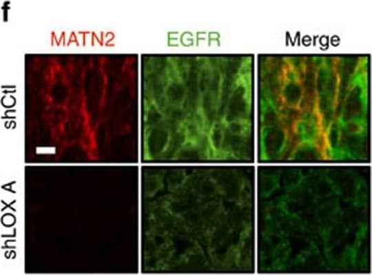
Detection of Mouse Matrilin-2 by Immunohistochemistry LOX controls EGF-dependent cell proliferation in vitro and tumour growth in vivo.(a,b) Graphs showing cell proliferation in control (shCtl) and LOX-depleted (shLOX A,B) MDA-MB-231 (a) and U87 (b) cells grown in 10% FBS (serum) or serum-free medium (serum free) and treated with PBS, rhMATN2 (500 ng ml−1) and EGF (50 ng ml−1) as indicated. All data are represented as mean±s.d. from four independent experiments. **P<0.01, Student's t-test. (c) Kaplan–Meier survival plot for mice following tail vein injection of control (shCtl) or LOX-depleted (shLOX A) MDA-MB-231 cells. Seven mice were used in each group. (d) H&E staining of lungs from mice in c. Scale bar, 500 μm. (e) Quantification of lung deposits in mice. Data is represented as mean±s.d. from seven samples. **P<0.05, Mann–Whitney analysis. (f) Photomicrographs for MATN2 (red) and EGFR (green) in lung deposits. Scale bar, 10 μm. (g) Quantification of samples in f. Data is represented as mean±s.e.m. from six samples. **P<0.01, Student's t-test. Image collected and cropped by CiteAb from the following publication (https://www.nature.com/articles/ncomms14909), licensed under a CC-BY license. Not internally tested by R&D Systems.
 View Larger
Detection of Mouse Matrilin-2 by Immunohistochemistry Chemical inhibition of LOX blocks tumour growth and metastasis.(a) Tumour growth in vehicle or CCT365623-treated MMTV-PyMT mice. Data are represented as mean±s.d. from six mice per group. **P<0.01, Student's t-test. (b) H&E stained lungs from vehicle or CCT365623 treated MMTV-PyMT mice. Scale bar, 100 μm. (c) Quantification of lung metastasis in mice from a. All data are presented as mean±range, n=6 animals. **P<0.05, Mann–Whitney analysis. (d) Tumour growth in water or BAPN-treated MMTV-PyMT mice. Data are represented as mean±s.d. from seven mice per group. **P<0.01, Student's t-test. (e) H&E stained lungs from water or BAPN-treated MMTV-PyMT mice. Scale bar, 100 μm. (f) Quantification of lung metastasis in mice from d. All data are presented as mean±range, n=7 animals. **P<0.05, Mann–Whitney analysis. (g,h) Confocal photomicrographs of MATN2 (red) and EGFR (green) staining in primary and metastatic tumours from vehicle, CCT365623, water or BAPN-treated MMTV-PyMT mice. Scale bars, 20 μm. (i–l) Quantification of MATN2 and EGFR staining in primary or metastatic tumours from g and h. All data are represented as mean±s.e.m. Samples from six vehicle or CCT365623 and seven water or BAPN-treated mice were analysed. **P<0.01, Student's t-test. Image collected and cropped by CiteAb from the following publication (https://www.nature.com/articles/ncomms14909), licensed under a CC-BY license. Not internally tested by R&D Systems.
Reconstitution Calculator
Preparation and Storage
- 12 months from date of receipt, -20 to -70 °C as supplied.
- 1 month, 2 to 8 °C under sterile conditions after reconstitution.
- 6 months, -20 to -70 °C under sterile conditions after reconstitution.
Background: Matrilin-2
Matrilin-2 is an extracellular matrix protein that belongs to the superfamily of von Willebrand factor A (VWA) containing proteins. It is expressed in many tissues and functions as a bridging component between other matrix proteins (1‑4). The human Matrilin-2 cDNA encodes a 956 amino acid (aa) precursor with a 23 aa signal sequence, two VWA domains separated by ten tandem EGF-like repeats, and a C-terminal coiled-coil domain (5, 6). Alternate splicing generates Isoform 2 (with an 18 aa deletion near the C-terminus), Isoform 3 (with a deletion of the fourth EGF-like repeat), and Isoform 4 (with a deletion of the first VWA and first EGF-like repeat). Human Matrilin-2 shares 87% and 84% aa sequence identity with mouse and canine Matrilin-2, respectively, and 27%, 22%, and 33% aa sequence identity with human Matrilin-1, -3, and -4, respectively. Matrilin-2 forms a variety of disulfide-linked oligomers via its coiled-coil domain (4, 7, 8, 9). It can assemble into homotrimers or heterotrimers with Matrilin-1 and/or Matrilin-4 (4, 7, 8) but has not been detected in heterotrimers containing Matrilin-3 (8). The VWA domains are thought to mediate Matrilin-Matrilin interactions as well as interactions with other matrix proteins such as Fibronectin, Collagen I, Fibrillin-2, and Laminin-1/Nidogen-1 complexes (7). Matrilin-2 knockout mice do not display any obvious abnormalities, suggesting that the expression of other molecules can compensate for the lack of Matrilin-2 (10).
- Wagener, R. et al. (2005) FEBS Lett. 579:3323.
- Deak, F. et al. (1999) Matrix Biol. 18:55.
- Whittaker, C.A. and R.O. Hynes (2002) Mol. Biol. Cell 13:3369.
- Piecha, D. et al. (1999) J. Biol. Chem. 274:13353.
- Muratoglu, S. et al. (2000) Cytogenet. Cell Genet. 90:323.
- Deak, F. et al. (1997) J. Biol. Chem. 272:9268.
- Piecha, D. et al. (2002) Biochem. J. 367:715.
- Frank, S. et al. (2002) J. Biol. Chem. 277:19071.
- Pan, O.H. and K. Beck (1998) J. Biol. Chem. 273:14205.
- Mates, L. et al. (2004) Matrix Biol. 23:195.
Product Datasheets
FAQs
No product specific FAQs exist for this product, however you may
View all Antibody FAQsReviews for Human Matrilin-2 Antibody
There are currently no reviews for this product. Be the first to review Human Matrilin-2 Antibody and earn rewards!
Have you used Human Matrilin-2 Antibody?
Submit a review and receive an Amazon gift card.
$25/€18/£15/$25CAN/¥75 Yuan/¥2500 Yen for a review with an image
$10/€7/£6/$10 CAD/¥70 Yuan/¥1110 Yen for a review without an image
