Human/Mouse Cyclin B1 Antibody Summary
Met1-Pro91
Accession # P14635
Applications
Please Note: Optimal dilutions should be determined by each laboratory for each application. General Protocols are available in the Technical Information section on our website.
Scientific Data
 View Larger
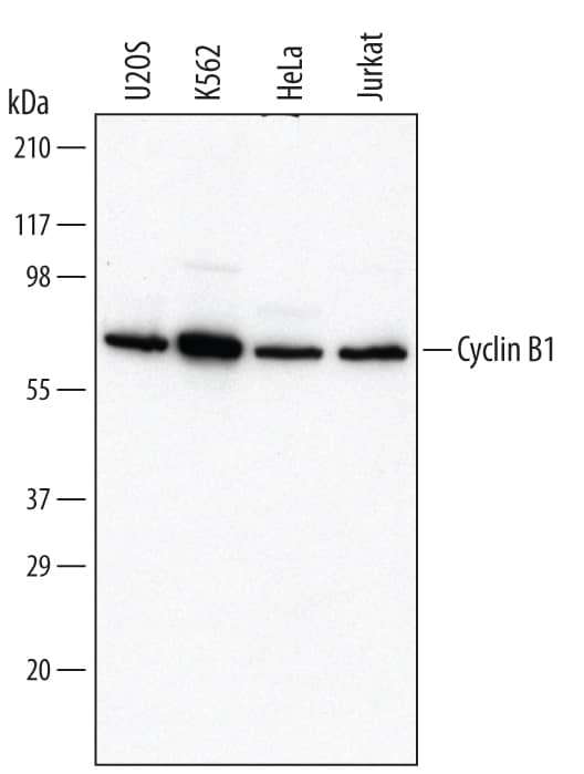
Detection of Human Cyclin B1 by Western Blot. Western blot shows lysates of U2OS human osteosarcoma cell line, K562 human chronic myelogenous leukemia cell line, HeLa human cervical epithelial carcinoma cell line, and Jurkat human acute T cell leukemia cell line. PVDF Membrane was probed with 0.2 µg/mL of Goat Anti-Human/Mouse Cyclin B1 Antigen Affinity-purified Polyclonal Antibody (Catalog # AF6000) followed by HRP-conjugated Anti-Goat IgG Secondary Antibody (Catalog # HAF017). A specific band was detected for Cyclin B1 at approximately 62 kDa (as indicated). This experiment was conducted under reducing conditions and using Immunoblot Buffer Group 1.
 View Larger
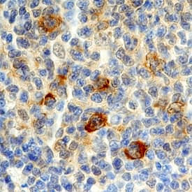
Cyclin B1 in Human Lymphoma. Cyclin B1 was detected in immersion fixed paraffin-embedded sections of human lymphoma using Goat Anti-Human/Mouse Cyclin B1 Antigen Affinity-purified Polyclonal Antibody (Catalog # AF6000) at 10 µg/mL overnight at 4 °C. Before incubation with the primary antibody, tissue was subjected to heat-induced epitope retrieval using Antigen Retrieval Reagent-Basic (Catalog # CTS013). Tissue was stained using the Anti-Goat HRP-DAB Cell & Tissue Staining Kit (brown; Catalog # CTS008) and counter-stained with hematoxylin (blue). Specific staining was localized to cytoplasm in lymphocytes. View our protocol for Chromogenic IHC Staining of Paraffin-embedded Tissue Sections.
 View Larger
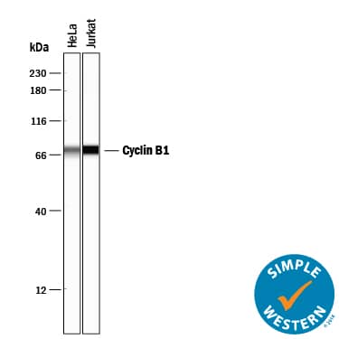
Detection of Human Cyclin B1 by Simple WesternTM. Simple Western lane view shows lysates of HeLa human cervical epithelial carcinoma cell line and Jurkat human acute T cell leukemia cell line, loaded at 0.2 mg/mL. A specific band was detected for Cyclin B1 at approximately 67 kDa (as indicated) using 2 µg/mL of Goat Anti-Human/Mouse Cyclin B1 Antigen Affinity-purified Polyclonal Antibody (Catalog # AF6000) followed by 1:50 dilution of HRP-conjugated Anti-Goat IgG Secondary Antibody (Catalog # HAF109). This experiment was conducted under reducing conditions and using the 12-230 kDa separation system.
 View Larger
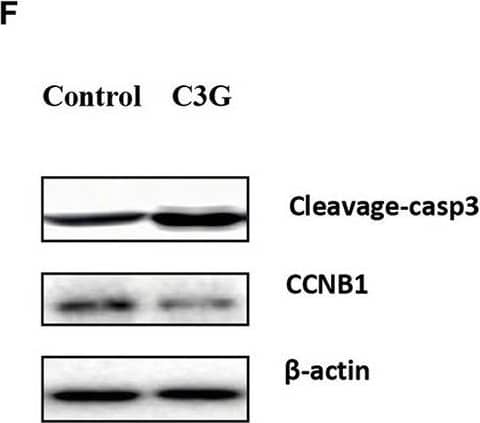
Detection of Mouse Cyclin B1 by Western Blot C3G inhibits the growth of B16-F10 cells in vivo. In situ tumor growth monitored via Xenogen IVIS imaging at different time points after implanting syngeneic B16-F10-luc tumors into male C57BL/6 mice (n = 10) either fed with control or C3G diet for 4 weeks. (A) Melanoma tumor growth was monitored in real-time via bioluminescent imaging of luciferase activity in live mice using the cryogenically cooled IVIS-imaging system from baseline to week 4 post implantation. (B) Graphical representation of tumor weight and tumor growth was monitored using Vernier calipers. (C) Photographic images of excised tumors were captured (n = 3). The largest tumor size in diameter is 2.0 cm. (D) Hematoxylin and eosin staining of tumor and capillaries are red (arrows). (E) Cleaved-caspase-3 was brownish in the cytoplasm (arrows) of the tumor and (F) Western blot analysis of caspase 3 and CCNB1 in C3G-treated mice tumors. (G), TUNEL staining to detect late apoptotic cells of the tumor (left panel) and C3G-treated mice tumors (Right panel). Late apoptotic cells had brownish nuclear regions (arrows). Differences with *p < 0.05, **p < 0.01, or ***p < 0.001 were considered statistically significant. Scale bar is 100 μm. Image collected and cropped by CiteAb from the following publication (https://pubmed.ncbi.nlm.nih.gov/31696058), licensed under a CC-BY license. Not internally tested by R&D Systems.
Preparation and Storage
- 12 months from date of receipt, -20 to -70 °C as supplied.
- 1 month, 2 to 8 °C under sterile conditions after reconstitution.
- 6 months, -20 to -70 °C under sterile conditions after reconstitution.
Background: Cyclin B1
Cyclin B1 (also CCNB1 and G2/mitotic-specific cyclin-B1) is a member of the cyclin AB subfamily, cyclin family of proteins. Although its predicted MW is 50 kDa, it runs anomalously at 62 kDa in SDS-PAGE. Cyclin B1 associates with both CDK1 and 2 providing substrate specificity to a phosphorylating complex. A phosphor‑CDK1:Cyclin B1 complex is inactive and cytosolic during interphase. At the beginning of mitosis, CDK1 is dephosphorylated and activated, and the CDK1:Cyclin B1 complex initiates formation of the mitotic scaffold. Human Cyclin B1 is 433 amino acids (aa) in length. It contains two cyclin box folds (aa 201‑290 and 298‑383) and two substrate binding sites (aa 298‑342 and 343‑380). Phosphorylation occurs at Ser9, Ser35, Ser69, and Thr321. There is one potential alternative start site at Met252 and deletions of aa 363‑399 and 365‑433. Over aa 1‑91, human Cyclin B1 shares 63% aa identity with mouse Cyclin B1.
Product Datasheets
Citations for Human/Mouse Cyclin B1 Antibody
R&D Systems personnel manually curate a database that contains references using R&D Systems products. The data collected includes not only links to publications in PubMed, but also provides information about sample types, species, and experimental conditions.
12
Citations: Showing 1 - 10
Filter your results:
Filter by:
-
Optoribogenetic control of regulatory RNA molecules
Authors: Sebastian Pilsl, Charles Morgan, Moujab Choukeife, Andreas Möglich, Günter Mayer
Nature Communications
-
A novel mouse model of CMT1B identifies hyperglycosylation as a new pathogenetic mechanism
Authors: Veneri FA, Prada V, Mastrangelo R et al.
Human molecular genetics
-
Liver X receptor activation reduces angiogenesis by impairing lipid raft localization and signaling of vascular endothelial growth factor receptor-2.
Authors: Noghero A, Perino A, Seano G, Saglio E, Lo Sasso G, Veglio F, Primo L, Hirsch E, Bussolino F, Morello F
Arterioscler Thromb Vasc Biol, 2012-06-21;32(9):2280-8.
-
Knockdown of Annexin-A1 Inhibits Growth, Migration and Invasion of Glioma Cells by Suppressing the PI3K/Akt Signaling Pathway
Authors: Liqing Wei, Li Li, Li Liu, Ru Yu, Xing Li, Zhenzhao Luo
ASN Neuro
-
Recombinant Human Cytomegalovirus Expressing an Analog-Sensitive Kinase pUL97 as Novel Tool for Functional Analyses
Authors: N Krämer, M Schütz, UG Mato, L Herhaus, M Marschall, C Zimmermann
Viruses, 2022-10-17;14(10):.
Species: Human
Sample Types: Cell Lysate
Applications: Bioassay -
Highly Conserved Interaction Profiles between Clinically Relevant Mutants of the Cytomegalovirus CDK-like Kinase pUL97 and Human Cyclins: Functional Significance of Cyclin H
Authors: M Schütz, R Müller, E Socher, C Wangen, F Full, E Wyler, D Wong, M Scherer, T Stamminger, S Chou, WD Rawlinson, ST Hamilton, H Sticht, M Marschall
International Journal of Molecular Sciences, 2022-10-05;23(19):.
Species: Human
Sample Types: Cell Lysates, Whole Cells
Applications: IP, Western Blot -
Metabolite and thymocyte development defects in ADA-SCID mice receiving enzyme replacement therapy
Authors: FA Moretti, G Giardino, TCH Attenborou, AS Gkazi, BK Margetts, G la Marca, L Fairbanks, T Crompton, HB Gaspar
Scientific Reports, 2021-12-01;11(1):23221.
Species: Mouse
Sample Types: Cell Lysates
Applications: Western Blot -
Functional Relevance of the Interaction between Human Cyclins and the Cytomegalovirus-Encoded CDK-Like Protein Kinase pUL97
Authors: M Schütz, M Steingrube, E Socher, R Müller, S Wagner, M Kögel, H Sticht, M Marschall
Viruses, 2021-06-27;13(7):.
Species: Human
Sample Types: Whole Cells
Applications: Immunoprecipitation -
Enhanced Expression of the Key Mitosis Regulator Cyclin B1 Is Mediated by PDZ-Binding Kinase in Islets of Pregnant Mice
Authors: T Uesato, T Ogihara, A Hara, H Iida, T Miyatsuka, Y Fujitani, S Takeda, H Watada
J Endocr Soc, 2018-01-30;2(3):207-219.
Species: Mouse
Sample Types: Cell Lysates
Applications: Western Blot -
Jab1 regulates Schwann cell proliferation and axonal sorting through p27.
Authors: Porrello, Emanuela, Rivellini, Cristina, Dina, Giorgia, Triolo, Daniela, Del Carro, Ubaldo, Ungaro, Daniela, Panattoni, Martina, Feltri, Maria La, Wrabetz, Lawrence, Pardi, Ruggero, Quattrini, Angelo, Previtali, Stefano
J Exp Med, 2013-12-16;211(1):29-43.
Species: Mouse
Sample Types: Tissue Homogenates
Applications: Western Blot -
Temporally distinct post-replicative repair mechanisms fill PRIMPOL-dependent ssDNA gaps in human cells
Authors: Tirman S, Quinet A, Wood M Et al.
Molecular cell
-
Cyanidin-3-o-glucoside pharmacologically inhibits tumorigenesis via estrogen receptor beta in melanoma mice
Authors: Liu M, Li H, Wang L et al.
Front Oncol
FAQs
No product specific FAQs exist for this product, however you may
View all Antibody FAQsReviews for Human/Mouse Cyclin B1 Antibody
There are currently no reviews for this product. Be the first to review Human/Mouse Cyclin B1 Antibody and earn rewards!
Have you used Human/Mouse Cyclin B1 Antibody?
Submit a review and receive an Amazon gift card.
$25/€18/£15/$25CAN/¥75 Yuan/¥2500 Yen for a review with an image
$10/€7/£6/$10 CAD/¥70 Yuan/¥1110 Yen for a review without an image

