Human/Mouse Pax3/Pax7 /Pax7 Antibody Summary
Met1-Ser215
Accession # NP_000429
*Small pack size (-SP) is supplied either lyophilized or as a 0.2 µm filtered solution in PBS.
Applications
Please Note: Optimal dilutions should be determined by each laboratory for each application. General Protocols are available in the Technical Information section on our website.
Scientific Data
 View Larger
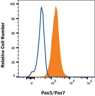
Detection of Pax3/Pax7 in B16-F1 cells by Flow Cytometry B16-F1 cells were stained with Mouse Anti-Human/Mouse Pax3/Pax7 /Pax7 Monoclonal Antibody (Catalog # MAB2457, filled histogram) or isotype control antibody (Catalog # MAB003, open histogram) followed by Allophycocyanin-conjugated Anti-Mouse IgG Secondary Antibody (Catalog # F0101B). To facilitate intracellular staining, cells were fixed with Flow Cytometry Fixation Buffer (Catalog # FC004) and permeabilized with Flow Cytometry Permeabilization/Wash Buffer I (Catalog # FC005). View our protocol for Staining Intracellular Molecules.
 View Larger
Pax3/Pax7 in B16‑F1 Mouse Cell Line. Pax3/Pax7 was detected in immersion fixed B16-F1 mouse melanoma cell line using Mouse Anti-Human/Mouse Pax3/Pax7 Monoclonal Antibody (Catalog # MAB2457) at 2 µg/mL for 3 hours at room temperature. Cells were stained using the NorthernLights™ 557-conjugated Anti-Mouse IgG Secondary Antibody (red; Catalog # NL007) and counterstained with DAPI (blue). Specific staining was localized to nuclei. View our protocol for Fluorescent ICC Staining of Cells on Coverslips.
 View Larger
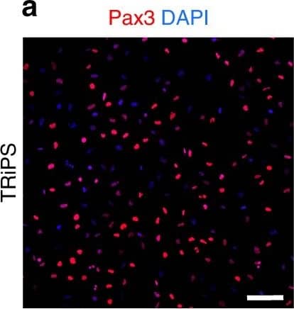
Detection of Human Pax3/Pax7 by Immunocytochemistry/Immunofluorescence Characterization of iMPCs during monolayer differentiation. a–e Representative immunostaining of Pax3 (a), Myf5 (b), MyoD (c), and MyoG (d), and corresponding quantification (e) during iMPC expansion. Scale bar=100 µm. f Representative FACS analysis for CD56 in H9 and TRiPSC derived iMPCs. g Representative immunostaining (top) and quantification (bottom) of Pax7+ and MyoG+ cell populations for H9 and TRiPS derived myotubes at 2 weeks of monolayer differentiation. (n = 6 samples from 2 differentiations for each cell line). h Representative immunostaining and quantification of GFP+/Pax7+ and GFP-/Pax7+ cell pools at 2 weeks of monolayer differentiation. Scale bar=50 µm. (n = 4 samples from 2 differentiations for each cell line). i Representative immunostaining and quantification of myotube diameter at 1, 2, and 4 weeks of monolayer differentiation. (*P < 0.05 vs. 1 week, #P < 0.05 vs. 4 week, Tukey–Kramer HSD test; n = 6 samples from 2 differentiations for each cell line). Scale bars=50 µm. Data are presented as mean ± SEM Image collected and cropped by CiteAb from the following publication (https://pubmed.ncbi.nlm.nih.gov/29317646), licensed under a CC-BY license. Not internally tested by R&D Systems.
 View Larger
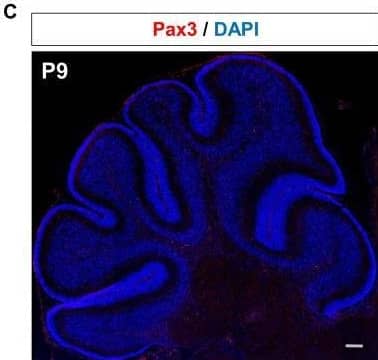
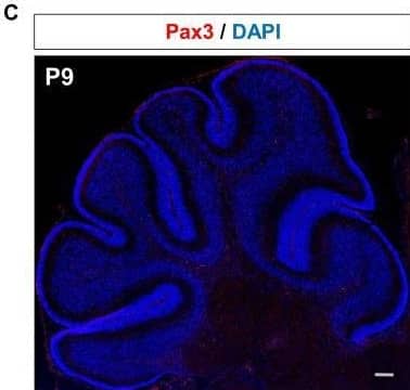
Detection of Pax3/Pax7/Pax7 by Immunohistochemistry Immunofluorescent analysis of Pax3 expression in the developing mouse cerebellum.(A) Bar plot showing the percentage of Pax3 + cells co-stained (y-axis) with cerebellar cell markers Pax2, Foxp2 and Calbindin at E12, E15, and P3 (x-axis). (B) Top: Immunofluorescent co-staining of Pax3 (red) and Foxp2 (green) in embryonic cerebellum at E15. Merged image is a composite image of the Pax3, Foxp2, and DAPI. Bottom: Immunofluorescent co-staining of Pax3 (red) and Calb (green) in the postnatal cerebellum at P0. Merged image is a composite image of the Pax3, Foxp2, and DAPI. Labels: VZ: Ventricular zone, EGL: External granular layer, PCL: Purkinje cell layer, ML: Molecular layer, Scalebars = 100 µm. (C) Immunofluorescent staining of Pax3 (red) in the developing cerebellum at P9. Image collected and cropped by CiteAb from the following open publication (https://pubmed.ncbi.nlm.nih.gov/35942939), licensed under a CC-BY license. Not internally tested by R&D Systems.
Reconstitution Calculator
Preparation and Storage
- 12 months from date of receipt, -20 to -70 °C as supplied.
- 1 month, 2 to 8 °C under sterile conditions after reconstitution.
- 6 months, -20 to -70 °C under sterile conditions after reconstitution.
Background: Pax3/Pax7
Pax3 belongs to the family of paired box transcription factors. Pax family proteins typically contain a paired box domain and a paired-type homeodomain. These transcription factors play critical roles during fetal development. Human and mouse Pax3 share 98% amino acid sequence identity.
Product Datasheets
Citations for Human/Mouse Pax3/Pax7 /Pax7 Antibody
R&D Systems personnel manually curate a database that contains references using R&D Systems products. The data collected includes not only links to publications in PubMed, but also provides information about sample types, species, and experimental conditions.
19
Citations: Showing 1 - 10
Filter your results:
Filter by:
-
Entinostat as a combinatorial therapeutic for rhabdomyosarcoma
Authors: Chauhan, S;Lian, E;Habib, I;Liu, Q;Anders, NM;Bugg, MM;Federman, NC;Reid, JM;Stewart, CF;Cates, T;Michalek, JE;Keller, C;
Scientific reports
Species: Human
Sample Types: Cell Lysates
Applications: Western Blot -
Transplantation of PSC-derived myogenic progenitors counteracts disease phenotypes in FSHD mice
Authors: Karim Azzag, Darko Bosnakovski, Sudheer Tungtur, Peter Salama, Michael Kyba, Rita C. R. Perlingeiro
npj Regenerative Medicine
-
Temporal analysis of enhancers during mouse cerebellar development reveals dynamic and novel regulatory functions
Authors: M Ramirez, Y Badayeva, J Yeung, J Wu, A Abdalla-Wy, E Yang, FANTOM 5 C, B Trost, SW Scherer, D Goldowitz
Elife, 2022-08-09;11(0):.
Species: Mouse
Sample Types: Whole Tissue
Applications: IHC -
Generation of Functional Brown Adipocytes from Human Pluripotent Stem Cells via Progression through a Paraxial Mesoderm State
Authors: L Zhang, J Avery, A Yin, AM Singh, TS Cliff, H Yin, S Dalton
Cell Stem Cell, 2020-08-11;0(0):.
Species: Human
Sample Types: Organoid, Whole Cells
Applications: Flow Cytometry, ICC, IHC -
Case report for an adolescent with germline RET mutation and alveolar rhabdomyosarcoma
Authors: Kenneth A. Crawford, Noah E. Berlow, Jennifer Tsay, Michael Lazich, Maria Mancini, Christopher Noakes et al.
Cold Spring Harb Mol Case Stud
-
Paraxial Mesoderm Is the Major Source of Lymphatic Endothelium
Authors: Oliver A. Stone, Didier Y.R. Stainier
Developmental Cell
-
The HDAC3–SMARCA4–miR-27a axis promotes expression of the PAX3:FOXO1 fusion oncogene in rhabdomyosarcoma
Authors: Narendra Bharathy, Noah E. Berlow, Eric Wang, Jinu Abraham, Teagan P. Settelmeyer, Jody E. Hooper et al.
Science Signaling
-
The SS18-SSX Fusion Oncoprotein Hijacks BAF Complex Targeting and Function to Drive Synovial Sarcoma
Authors: Matthew J. McBride, John L. Pulice, Hannah C. Beird, Davis R. Ingram, Andrew R. D’Avino, Jack F. Shern et al.
Cancer Cell
-
Methotrexate and Valproic Acid Affect Early Neurogenesis of Human Amniotic Fluid Stem Cells from Myelomeningocele
Authors: V Sahakyan, E Pozzo, R Duelen, J Deprest, M Sampaolesi
Stem Cells Int, 2017-09-13;2017(0):6101609.
Species: Human
Sample Types: Whole Cells
Applications: ICC -
Mesodermal iPSC–derived progenitor cells functionally regenerate cardiac and skeletal muscle
Authors: Mattia Quattrocelli, Melissa Swinnen, Giorgia Giacomazzi, Jordi Camps, Ines Barthélemy, Gabriele Ceccarelli et al.
Journal of Clinical Investigation
-
microRNA-155, induced by interleukin-1ss, represses the expression of microphthalmia-associated transcription factor (MITF-M) in melanoma cells.
Authors: Arts N, Cane S, Hennequart M, Lamy J, Bommer G, Van den Eynde B, De Plaen E
PLoS ONE, 2015-04-08;10(4):e0122517.
Species: Human
Sample Types: Cell Lysates
Applications: Western Blot -
Derivation and expansion of PAX7-positive muscle progenitors from human and mouse embryonic stem cells.
Authors: Shelton M, Metz J, Liu J, Carpenedo R, Demers S, Stanford W, Skerjanc I
Stem Cell Reports, 2014-08-07;3(3):516-29.
Species: Human, Mouse
Sample Types: Cell Culture Supernates
Applications: IHC -
Lineage of origin in rhabdomyosarcoma informs pharmacological response.
Authors: Abraham J, Nunez-Alvarez Y, Hettmer S, Carrio E, Chen H, Nishijo K, Huang E, Prajapati S, Walker R, Davis S, Rebeles J, Wiebush H, McCleish A, Hampton S, Bjornson C, Brack A, Wagers A, Rando T, Capecchi M, Marini F, Ehler B, Zarzabal L, Goros M, Michalek J, Meltzer P, Langenau D, LeGallo R, Mansoor A, Chen Y, Suelves M, Rubin B, Keller C
Genes Dev, 2014-07-15;28(14):1578-91.
Species: Human, Mouse
Sample Types: Whole Cells
Applications: ICC -
Cell-cycle dependent expression of a translocation-mediated fusion oncogene mediates checkpoint adaptation in rhabdomyosarcoma.
Authors: Kikuchi K, Hettmer S, Aslam M, Michalek J, Laub W, Wilky B, Loeb D, Rubin B, Wagers A, Keller C
PLoS Genet, 2014-01-16;10(1):e1004107.
Species: Human, Mouse
Sample Types: Whole Cells
Applications: ICC -
Pax3 and Zic1 trigger the early neural crest gene regulatory network by the direct activation of multiple key neural crest specifiers.
Authors: Plouhinec J, Roche D, Pegoraro C, Figueiredo A, Maczkowiak F, Brunet L, Milet C, Vert J, Pollet N, Harland R, Monsoro-Burq A
Dev Biol, 2013-12-17;386(2):461-72.
Species: Human
Sample Types: Whole Cells
Applications: Functional Assay -
Functional dissection of Pax3 in paraxial mesoderm development and myogenesis.
Authors: Magli A, Schnettler E, Rinaldi F, Bremer P, Perlingeiro R
Stem Cells, 2013-01-01;31(1):59-70.
Species: Mouse
Sample Types: Cell Lysates
Applications: Western Blot -
Selective development of myogenic mesenchymal cells from human embryonic and induced pluripotent stem cells.
Authors: Awaya, Tomonari, Kato, Takeo, Mizuno, Yuta, Chang, Hsi, Niwa, Akira, Umeda, Katsutsu, Nakahata, Tatsutos, Heike, Toshio
PLoS ONE, 2012-12-07;7(12):e51638.
Species: Human
Sample Types: Whole Cells
Applications: ICC -
MyoD directly up-regulates premyogenic mesoderm factors during induction of skeletal myogenesis in stem cells.
Authors: Gianakopoulos PJ, Mehta V, Voronova A, Cao Y, Yao Z, Coutu J, Wang X, Waddington MS, Tapscott SJ, Skerjanc IS
J. Biol. Chem., 2010-11-15;286(4):2517-25.
Species: Mouse
Sample Types: Cell Lysates
Applications: Western Blot -
Pax3 activation promotes the differentiation of mesenchymal stem cells toward the myogenic lineage.
Authors: Gang EJ, Bosnakovski D, Simsek T, To K, Perlingeiro RC
Exp. Cell Res., 2008-03-05;314(8):1721-33.
Species: Mouse
Sample Types: Cell Lysates
Applications: Western Blot
FAQs
No product specific FAQs exist for this product, however you may
View all Antibody FAQsReviews for Human/Mouse Pax3/Pax7 /Pax7 Antibody
Average Rating: 5 (Based on 1 Review)
Have you used Human/Mouse Pax3/Pax7 /Pax7 Antibody?
Submit a review and receive an Amazon gift card.
$25/€18/£15/$25CAN/¥75 Yuan/¥2500 Yen for a review with an image
$10/€7/£6/$10 CAD/¥70 Yuan/¥1110 Yen for a review without an image
Filter by:

