Human/Mouse/Rat FABP1/L-FABP Antibody Summary
Met1-Ile127
Accession # AAA52418
Applications
Please Note: Optimal dilutions should be determined by each laboratory for each application. General Protocols are available in the Technical Information section on our website.
Scientific Data
 View Larger
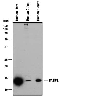
Detection of Human FABP1/L‑FABP by Western Blot. Western blot shows lysates of human liver tissue, human colon tissue, and human kidney tissue. PVDF membrane was probed with 0.25 µg/mL of Mouse Anti-Human/Mouse/Rat FABP1/L-FABP Monoclonal Antibody (Catalog # MAB2964) followed by HRP-conjugated Anti-Mouse IgG Secondary Antibody (Catalog # HAF018). A specific band was detected for FABP1/ L-FABP at approximately 14 kDa (as indicated). This experiment was conducted under reducing conditions and using Immunoblot Buffer Group 1.
 View Larger
Detection of Human, Mouse, and Rat FABP1/L‑FABP by Western Blot. Western blot shows lysates of HepG2 human hepatocellular carcinoma cell line, mouse liver tissue, and rat liver tissue. PVDF membrane was probed with 0.25 µg/mL of Mouse Anti-Human/Mouse/Rat FABP1/ L-FABP Monoclonal Antibody (Catalog # MAB2964) followed by HRP-conjugated Anti-Mouse IgG Secondary Antibody (Catalog # HAF018). A specific band was detected for FABP1/L-FABP at approximately 14 kDa (as indicated). This experiment was conducted under reducing conditions and using Immunoblot Buffer Group 1.
 View Larger
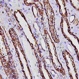
FABP1/L‑FABP in Human Kidney. FABP1/L-FABP was detected in immersion fixed paraffin-embedded sections of human kidney using Mouse Anti-Human/Mouse/Rat FABP1/L-FABP Monoclonal Antibody (Catalog # MAB2964) at 15 µg/mL overnight at 4 °C. Tissue was stained using the Anti-Mouse HRP-DAB Cell & Tissue Staining Kit (brown; Catalog # CTS002) and counterstained with hematoxylin (blue). Specific staining was localized to convoluted tubules. View our protocol for Chromogenic IHC Staining of Paraffin-embedded Tissue Sections.
 View Larger
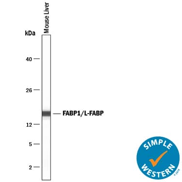
Detection of Mouse FABP1/L‑FABP by Simple WesternTM. Simple Western lane view shows lysates of mouse liver tissue, loaded at 0.2 mg/mL. A specific band was detected for FABP1/L‑FABP at approximately 16 kDa (as indicated) using 5 µg/mL of Mouse Anti-Human/Mouse/Rat FABP1/ L‑FABP Monoclonal Antibody (Catalog # MAB2964). This experiment was conducted under reducing conditions and using the 12-230 kDa separation system.
Reconstitution Calculator
Preparation and Storage
- 12 months from date of receipt, -20 to -70 °C as supplied.
- 1 month, 2 to 8 °C under sterile conditions after reconstitution.
- 6 months, -20 to -70 °C under sterile conditions after reconstitution.
Background: FABP1/L-FABP
FABP1, also known as liver FABP (L-FABP, Z-protein and squalene-and sterol-carrier protein [SCP]) is a member of the intracellular FABP family. It is highly expressed in the liver, intestine, kidney and lung. FABP1 binds free fatty acids and their co-enzyme A derivatives and may be involved in intracellular lipid transport.
Product Datasheets
FAQs
No product specific FAQs exist for this product, however you may
View all Antibody FAQsReviews for Human/Mouse/Rat FABP1/L-FABP Antibody
Average Rating: 4 (Based on 4 Reviews)
Have you used Human/Mouse/Rat FABP1/L-FABP Antibody?
Submit a review and receive an Amazon gift card.
$25/€18/£15/$25CAN/¥75 Yuan/¥2500 Yen for a review with an image
$10/€7/£6/$10 CAD/¥70 Yuan/¥1110 Yen for a review without an image
Filter by:
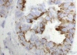
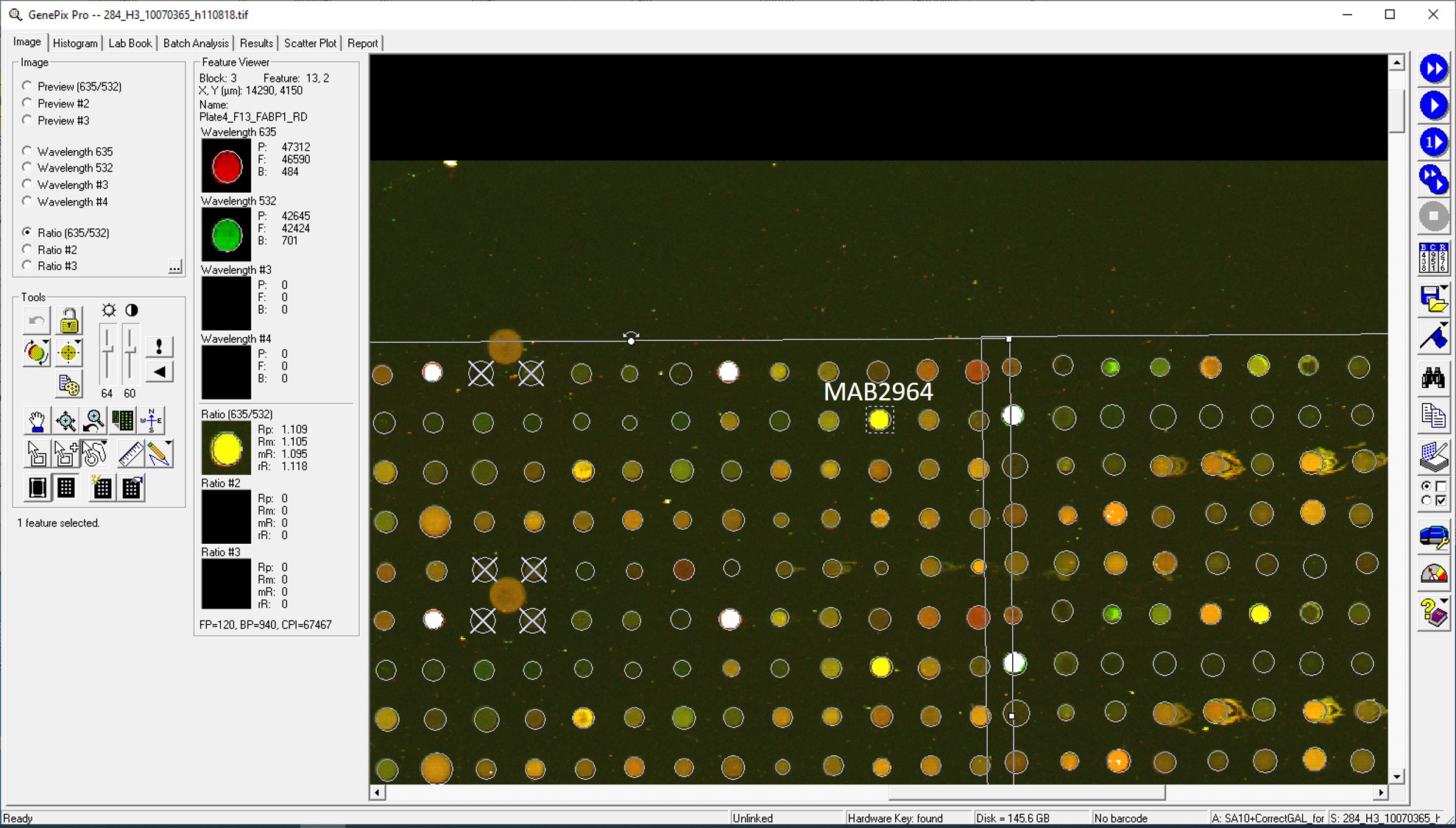
Antibody was printed on custom arrays and incubated with fluorescently labeled human EDTA plasma
