Human/Mouse/Rat GAD1/GAD67 Antibody Summary
Ala2-Asn97
Accession # Q99259
Applications
Please Note: Optimal dilutions should be determined by each laboratory for each application. General Protocols are available in the Technical Information section on our website.
Scientific Data
 View Larger
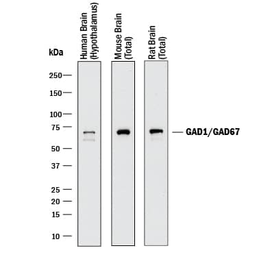
Detection of Human, Mouse, and Rat GAD1/GAD67 by Western Blot. Western blot shows lysates of human brain (hypothalamus) tissue, mouse brain (total) tissue, and rat brain (total) tissue. PVDF membrane was probed with 1 µg/mL of Goat Anti-Human/Mouse/Rat GAD1/GAD67 Antigen Affinity-purified Polyclonal Antibody (Catalog # AF2086) followed by HRP-conjugated Anti-Goat IgG Secondary Antibody HAF017). A specific band was detected for GAD1/GAD67 at approximately 67 kDa (as indicated). This experiment was conducted under reducing conditions and using Immunoblot Buffer Group 1.
 View Larger
GAD1/GAD67 in Human Spinal Cord. GAD1/GAD67 was detected in immersion fixed paraffin-embedded sections of human spinal cord using 15 µg/mL Goat Anti-Human/Mouse/Rat GAD1/GAD67 Antigen Affinity-purified Polyclonal Antibody (Catalog # AF2086) overnight at 4 °C. Tissue was stained with the Anti-Goat HRP-DAB Cell & Tissue Staining Kit (brown; CTS008) and counterstained with hematoxylin (blue). View our protocol for Chromogenic IHC Staining of Paraffin-embedded Tissue Sections.
 View Larger
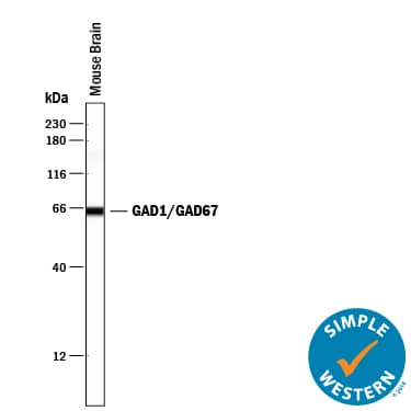
Detection of Mouse GAD1/GAD67 by Simple WesternTM. Simple Western lane view shows lysates of mouse brain tissue, loaded at 0.2 mg/mL. A specific band was detected for GAD1/GAD67 at approximately 65 kDa (as indicated) using 1 µg/mL of Goat Anti-Human/Mouse/Rat GAD1/GAD67 Antigen Affinity-purified Polyclonal Antibody (Catalog # AF2086) followed by 1:50 dilution of HRP-conjugated Anti-Goat IgG Secondary Antibody (HAF109). This experiment was conducted under reducing conditions and using the 12-230 kDa separation system.
Reconstitution Calculator
Preparation and Storage
- 12 months from date of receipt, -20 to -70 °C as supplied.
- 1 month, 2 to 8 °C under sterile conditions after reconstitution.
- 6 months, -20 to -70 °C under sterile conditions after reconstitution.
Background: GAD1/GAD67
GAD1, also named 67 kDa or brain GAD, is an enzyme that catalyzes the formation of the inhibitory neurotransmitter gamma -amino butyric acid (GABA) from glutamate. GAD1 is also expressed in multiple non-neuronal tissues during development.
Product Datasheets
Citations for Human/Mouse/Rat GAD1/GAD67 Antibody
R&D Systems personnel manually curate a database that contains references using R&D Systems products. The data collected includes not only links to publications in PubMed, but also provides information about sample types, species, and experimental conditions.
14
Citations: Showing 1 - 10
Filter your results:
Filter by:
-
Altered Parvalbumin Basket Cell Terminals in the Cortical Visuospatial Working Memory Network in Schizophrenia
Authors: Kenneth N. Fish, Brad R. Rocco, Adam M. DeDionisio, Samuel J. Dienel, Robert A. Sweet, David A. Lewis
Biological Psychiatry
-
Dorsal striatum c-Fos activity in perseverative ephrin-A2A5−/− mice and the cellular effect of low-intensity rTMS
Authors: Maitri Tomar, Jennifer Rodger, Jessica Moretti
Frontiers in Neural Circuits
-
Markedly Lower Glutamic Acid Decarboxylase 67 Protein Levels in a Subset of Boutons in Schizophrenia
Authors: Brad R. Rocco, David A. Lewis, Kenneth N. Fish
Biological Psychiatry
-
Restoration of brain circulation and cellular functions hours post-mortem
Authors: Zvonimir Vrselja, Stefano G. Daniele, John Silbereis, Francesca Talpo, Yury M. Morozov, André M. M. Sousa et al.
Nature
-
Ciliary neuropeptidergic signaling dynamically regulates excitatory synapses in postnatal neocortical pyramidal neurons
Authors: Lauren Tereshko, Ya Gao, Brian A Cary, Gina G Turrigiano, Piali Sengupta
eLife
-
Isolated loss of the AUTS2 long isoform, brain-wide or targeted to Calbindin -lineage cells, generates a specific suite of brain, behavioral and molecular pathologies
Authors: Song, Y;Seward, CH;Chen, CY;LeBlanc, A;Leddy, AM;Stubbs, L;
bioRxiv : the preprint server for biology
Species: Transgenic Mouse
Sample Types: Whole Tissue
Applications: Immunohistochemistry -
Spatially resolved transcriptomics reveals genes associated with the vulnerability of middle temporal gyrus in Alzheimer's disease
Authors: S Chen, Y Chang, L Li, D Acosta, Y Li, Q Guo, C Wang, E Turkes, C Morrison, D Julian, ME Hester, DW Scharre, C Santiskulv, SX Song, JT Plummer, GE Serrano, TG Beach, KE Duff, Q Ma, H Fu
Acta neuropathologica communications, 2022-12-21;10(1):188.
Species: Human
Sample Types: Whole Tissue
Applications: IHC -
The beta2V287L nicotinic subunit linked to sleep-related epilepsy differently affects fast-spiking and regular spiking somatostatin-expressing neurons in murine prefrontal cortex
Authors: S Meneghini, D Modena, G Colombo, A Coatti, N Milani, L Madaschi, A Amadeo, A Becchetti
Progress in neurobiology, 2022-05-02;0(0):102279.
Species: Mouse
Sample Types: Whole Tissue
Applications: IHC -
Regulation of prefrontal patterning and connectivity by retinoic acid
Authors: M Shibata, K Pattabiram, B Lorente-Ga, D Andrijevic, SK Kim, N Kaur, SK Muchnik, X Xing, G Santpere, AMM Sousa, N Sestan
Nature, 2021-10-01;0(0):.
Species: Human, Mouse
Sample Types: Whole Tissue
Applications: IHC -
Altered Parvalbumin Basket Cell Terminals in the Cortical Visuospatial Working Memory Network in Schizophrenia
Authors: Kenneth N. Fish, Brad R. Rocco, Adam M. DeDionisio, Samuel J. Dienel, Robert A. Sweet, David A. Lewis
Biological Psychiatry
Species: Human
Sample Types: Whole Tissue
Applications: Immunohistochemistry -
Alterations in a Unique Class of Cortical Chandelier Cell Axon Cartridges in Schizophrenia
Authors: Kenneth N Fish
Biol. Psychiatry, 2016-09-29;0(0):.
Species: Human
Sample Types: Whole Tissue
Applications: IHC -
Transporters MCT8 and OATP1C1 maintain murine brain thyroid hormone homeostasis.
Authors: Mayerl S, Muller J, Bauer R, Richert S, Kassmann C, Darras V, Buder K, Boelen A, Visser T, Heuer H
J Clin Invest, 2014-04-01;124(5):1987-99.
Species: Human
Sample Types: Whole Tissue
Applications: IHC -
Locomotor deficiencies and aberrant development of subtype-specific GABAergic interneurons caused by an unliganded thyroid hormone receptor alpha1.
Authors: Wallis K, Sjogren M, van Hogerlinden M, Silberberg G, Fisahn A, Nordstrom K, Larsson L, Westerblad H, Morreale de Escobar G, Shupliakov O, Vennstrom B
J. Neurosci., 2008-02-20;28(8):1904-15.
Species: Mouse
Sample Types: Whole Tissue
Applications: IHC-Fr -
The 7q11.23 Protein DNAJC30 Interacts with ATP Synthase and Links Mitochondria to Brain Development.
Authors: Tebbenkamp ATN, Varela L, Choi J et al.
Cell.
FAQs
No product specific FAQs exist for this product, however you may
View all Antibody FAQsReviews for Human/Mouse/Rat GAD1/GAD67 Antibody
Average Rating: 1 (Based on 1 Review)
Have you used Human/Mouse/Rat GAD1/GAD67 Antibody?
Submit a review and receive an Amazon gift card.
$25/€18/£15/$25CAN/¥75 Yuan/¥2500 Yen for a review with an image
$10/€7/£6/$10 CAD/¥70 Yuan/¥1110 Yen for a review without an image
Filter by:
Does not work with incubation at 1:500 to 1:1000 dilutions overnight plus secondary AlexaFluor (multiple tested). No fluorescence detected. Sample tissue fixed with PFA.


