Human/Mouse/Rat GDF-8/Myostatin Biotinylated Antibody Summary
Asp268-Ser376
Accession # O08689
Applications
Please Note: Optimal dilutions should be determined by each laboratory for each application. General Protocols are available in the Technical Information section on our website.
Scientific Data
 View Larger
GDF‑8/Myostatin in Mouse Embryonic Lung. GDF-8/Myostatin was detected in immersion fixed frozen sections of mouse embryo (lung) using Goat Anti-Human/Mouse/Rat GDF-8/Myostatin Biotinylated Antigen Affinity-purified Polyclonal Antibody (Catalog # BAF788) at 15 µg/mL overnight at 4 °C. Tissue was stained using the Anti-Goat HRP-DAB Cell & Tissue Staining Kit (brown; CTS008) and counterstained with hematoxylin (blue). View our protocol for Chromogenic IHC Staining of Frozen Tissue Sections.
 View Larger
Detection of GDF‑8/Myostatin in Mouse Embryo. GDF‑8/Myostatin was detected in immersion fixed paraffin-embedded 13 d.p.c. sections of Mouse Embryo using Goat Anti-Human/Mouse/Rat GDF‑8/Myostatin Biotinylated Antigen Affinity-purified Polyclonal Antibody (Catalog # BAF788) at 25 µg/mL for 1 hour at room temperature followed by incubation with the Anti-Goat IgG VisUCyte™ HRP Polymer Antibody (Catalog # VC004). Before incubation with the primary antibody, tissue was subjected to heat-induced epitope retrieval using VisUCyte Antigen Retrieval Reagent-Basic (Catalog # VCTS021). Tissue was stained using DAB (brown) and counterstained with hematoxylin (blue). Specific staining was localized to muscle cells in developing tongue. View our protocol for IHC Staining with VisUCyte HRP Polymer Detection Reagents.
Reconstitution Calculator
Preparation and Storage
- 12 months from date of receipt, -20 to -70 °C as supplied.
- 1 month, 2 to 8 °C under sterile conditions after reconstitution.
- 6 months, -20 to -70 °C under sterile conditions after reconstitution.
Background: GDF-8/Myostatin
Growth Differentiation Factor 8 (GDF-8), also known as myostatin, is a member of the TGF-beta superfamily that is expressed specifically in developing and adult skeletal muscle. GDF-8 cDNA encodes a 376 amino acid (aa) prepropeptide with a 24 aa residue signal peptide, a 223 aa residue amino-terminal propeptide, and a 109 aa residue carboxy-terminal mature protein. Mature GDF-8 contains the canonical 7-cysteine motif common to other TGF-beta superfamily members. Similar to the TGF‑ beta s, activins and BMP-11, GDF-8 also contains one extra pair of cysteine residues that is not found in other family members. The bioactive form of GDF-8 is a homodimer with an apparent molecular weight of approximately 25 kDa. GDF-8 is highly conserved across species. At the amino acid sequence level, mature human, mouse, rat and cow GDF-8 are 100% identical. Within the TGF-beta superfamily, GDF-8 is most closely related to BMP-11, a mammalian protein that acts as a dorsal mesoderm and neural inducer in Xenopus explants. The two proteins share 90% amino acid sequence identity within their mature chain. A targeted disruption of GDF-8 in mouse results in large mice with a widespread increase in skeletal muscle mass, indicating that GDF-8 is a negative regulator of skeletal muscle growth. A mutation in the bovine GDF-8 gene has been shown to be responsible for the double-muscled phenotype in cattle breeds such as Belgian Blue cattle that is characterized by an increase in muscle mass. GDF‑8 has also been shown to inhibit preadipocyte differentiation to adipocytes. Mature GDF-8 binds to activin type II receptors and the binding is antagonized by the activin-binding protein, follistatin. R&D Systems recombinant GDF-8 preparations have been shown to act similarly to Activin A in both the Xenopus animal cap and the K562 assays.
- Storm, E.E. et al. (1994) Nature 368:639
- Sharma, M. et al. (1999) J. Cell Physiol. 180:1
- McPherron, A.C. et al. (1997) Nature 387:83
- Lee, S.J. et al. (2001) Proc. Natl. Acad. Sci. USA 98:9306
- Kim, H.S. et al. (2001) Biochem. Biophys. Res. Commun. 281:902
Product Datasheets
Citations for Human/Mouse/Rat GDF-8/Myostatin Biotinylated Antibody
R&D Systems personnel manually curate a database that contains references using R&D Systems products. The data collected includes not only links to publications in PubMed, but also provides information about sample types, species, and experimental conditions.
3
Citations: Showing 1 - 3
Filter your results:
Filter by:
-
Quantification of GDF11 and Myostatin in Human Aging and Cardiovascular Disease
Authors: Marissa J Schafer
Cell Metab, 2016-06-14;23(6):1207-15.
-
Structural basis of specific inhibition of extracellular activation of pro- or latent myostatin by the monoclonal antibody SRK-015
Authors: KB Dagbay, E Treece, FC Streich, JW Jackson, RR Faucette, A Nikiforov, SC Lin, CJ Boston, SB Nicholls, AD Capili, GJ Carven
J. Biol. Chem., 2020-02-19;295(16):5404-5418.
Species: Human
Sample Types: Cell Culture Supernates
-
Myostatin as a mediator of sarcopenia versus homeostatic regulator of muscle mass: insights using a new mass spectrometry-based assay
Authors: H. Robert Bergen, Joshua N. Farr, Patrick M. Vanderboom, Elizabeth J. Atkinson, Thomas A. White, Ravinder J. Singh et al.
Skeletal Muscle
FAQs
No product specific FAQs exist for this product, however you may
View all Antibody FAQsReviews for Human/Mouse/Rat GDF-8/Myostatin Biotinylated Antibody
Average Rating: 1 (Based on 1 Review)
Have you used Human/Mouse/Rat GDF-8/Myostatin Biotinylated Antibody?
Submit a review and receive an Amazon gift card.
$25/€18/£15/$25CAN/¥75 Yuan/¥2500 Yen for a review with an image
$10/€7/£6/$10 CAD/¥70 Yuan/¥1110 Yen for a review without an image
Filter by:
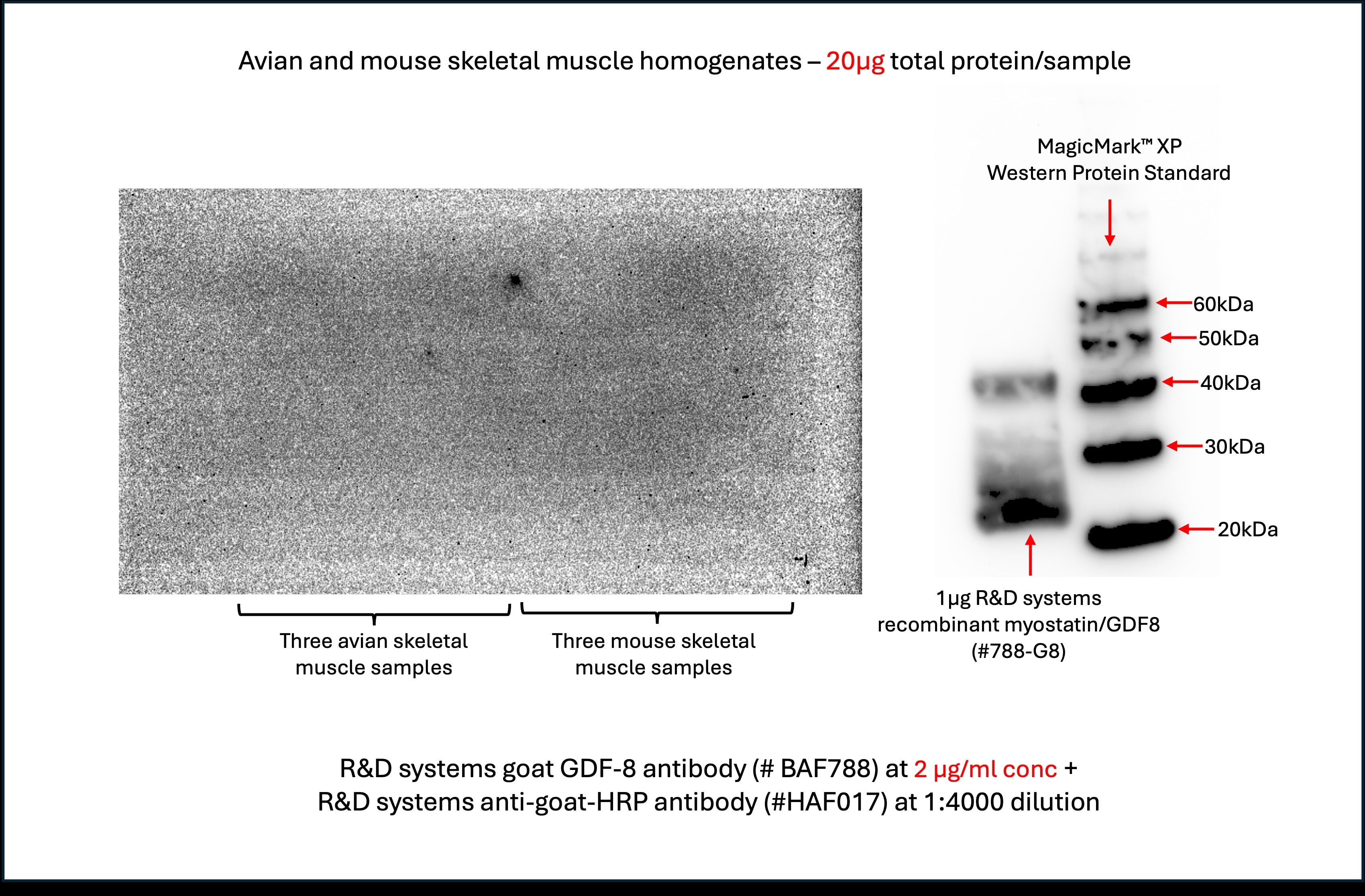
The test samples consisted of skeletal muscle tissue homogenates derived from mouse and avian muscle tissues. The homogenisation buffer contained Roche mini cOmplete protease inhibitor cocktail. The positive control used was 1μg of R&D systems recombinant myostatin/GDF8 (#788-G8).
I had loaded 20μg of total protein for each of the avian and mouse samples, along with1μg of R&D systems recombinant myostatin/GDF8 (#788-G8) into a Bio-Rad PROTEAN TGX 4-15% gel.
The electrophoresis was performed with NuPAGE MES running buffer at 90V constant current, and the proteins were subsequently blotted onto a 0.2μm nitrocellulose membrane. To check for transfer efficiency and protein integrity, i had stained the blot with Ponceau S and found that the transfer was efficient and the proteins were not degraded. Subsequently, i destained the blot with TBS-T and deionised water, and blocked the blots with 5% milk in TBS-T.
Prior to incubating the membrane in primary antibody, i had cut the membrane such that one section contained all the animal tissue samples and the other section contained the recombinant GDF-8/myostatin and the MagicMark XP western standard.
The cut sections were incubated separately with identical concentrations of primary and secondary antibodies - R&D systems goat polyclonal GDF8 antibody (BAF788) at 2 µg/mL concentration, and R&D system anti-goat secondary antibody (HAF017) at 1:4000 dilution.
The recombinant GDF-8/Myostatin positive control and the MagicMark XP western standards produced clear and robust signals.
However, the sections containing the samples derived from animal tissues (avian and mouse) failed to produce any discernible signal.
I had repeated the same experiment above three times with increasing concentrations of primary antibody - 0.1, 0.25 and 2 µg/mL. However, all three experiments yielded the same result - strong signal for the recombinant GDF-8/Myostatin, but no visible signal detected with the avian and mouse tissue samples.

