Human/Mouse/Rat Histone H2AX Antibody Summary
Gly3-Ser131
Accession # P16104
Applications
Please Note: Optimal dilutions should be determined by each laboratory for each application. General Protocols are available in the Technical Information section on our website.
Scientific Data
 View Larger
Detection of Human/Mouse/Rat Histone H2AX by Western Blot. Western blot shows lysates of HEK293 human embryonic kidney cell line, K562 human chronic myelogenous leukemia cell line, Balb/3T3 mouse embryonic fibroblast cell line, and Nb2-11 rat lymphoma cell line. PVDF membrane was probed with 0.5 µg/mL of Mouse Anti-Human/Mouse/Rat Histone H2AX Monoclonal Antibody (Catalog # MAB3406) followed by HRP-conjugated Anti-Mouse IgG Secondary Antibody (Catalog # HAF007). A specific band was detected for Histone H2AX at approximately 20 kDa (as indicated). This experiment was conducted under reducing conditions and using Immunoblot Buffer Group 1.
 View Larger
Histone H2AX in HeLa Human Cell Line. Histone H2AX was detected in immersion fixed HeLa human cervical epithelial carcinoma cell line using Mouse Anti-Human/Mouse/Rat Histone H2AX Monoclonal Antibody (Catalog # MAB3406) at 8 µg/mL for 3 hours at room temperature. Cells were stained using the Northern-Lights™ 557-conjugated Anti-Mouse IgG Secondary Antibody (red; Catalog # NL007) and counterstained with DAPI (blue). Specific staining was localized to nuclei. View our protocol for Fluorescent ICC Staining of Cells on Coverslips.
 View Larger
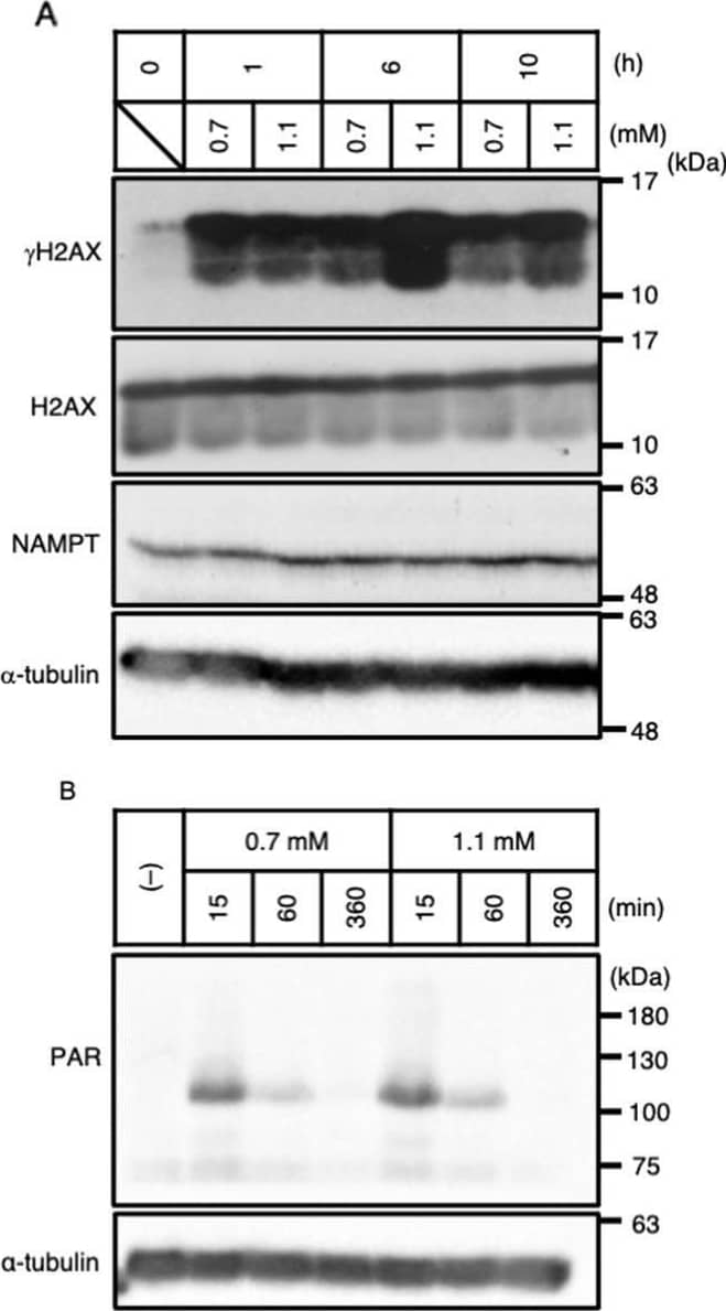
Detection of Histone H2AX by Western Blot The NAMPT-dependent NAD+ salvage pathway is necessary for the recovery of NAD+ & induction of apoptosis under weak H2O2 stimulation. A The amounts of endogenous gamma H2AX & NAMPT proteins under 0.7 & 1.1 mM H2O2 stimulation determined by immunoblotting (n = 3). Image collected & cropped by CiteAb from the following open publication (https://pubmed.ncbi.nlm.nih.gov/35410407), licensed under a CC-BY license. Not internally tested by R&D Systems.
Reconstitution Calculator
Preparation and Storage
- 12 months from date of receipt, -20 to -70 °C as supplied.
- 1 month, 2 to 8 °C under sterile conditions after reconstitution.
- 6 months, -20 to -70 °C under sterile conditions after reconstitution.
Background: Histone H2AX
Histone H2AX is a core histone protein that is phosphorylated at S139 in cells exposed to DNA double-strand break-inducing agents, such as ionizing radiation. The S139 phosphorylated H2AX, termed gamma -H2AX, marks the site of DNA double-strand breaks and serves to recruit cell cycle checkpoint and DNA repair factors to the site of damage.
Product Datasheets
Citations for Human/Mouse/Rat Histone H2AX Antibody
R&D Systems personnel manually curate a database that contains references using R&D Systems products. The data collected includes not only links to publications in PubMed, but also provides information about sample types, species, and experimental conditions.
11
Citations: Showing 1 - 10
Filter your results:
Filter by:
-
Hepatic AhR Activation by TCDD Induces Obesity and Steatosis via Hepatic Plasminogen Activator Inhibitor-1 (PAI-1)
Authors: Oh, SJ;Im, S;Kang, S;Lee, AG;Lee, BC;Pak, YK;
International journal of molecular sciences
Species: Mouse
Sample Types: Tissue Homogenates
Applications: Western Blot -
Mitoregulin Promotes Cell Cycle Progression in Non-Small Cell Lung Cancer Cells
Authors: Stein, CS;Linzer, CR;Heer, CD;Witmer, NH;Cochran, JD;Spitz, DR;Boudreau, RL;
International journal of molecular sciences
Species: Human
Sample Types: Cell Lysates
Applications: Western Blot -
Inflammasome Coordinates Senescent Chronic Wound Induced by Thalassophryne nattereri Venom
Authors: Lima, C;Andrade-Barros, AI;Carvalho, FF;Falc�o, MAP;Lopes-Ferreira, M;
International journal of molecular sciences
Species: Mouse
Sample Types: Whole Tissue
Applications: IHC -
NAMPT-dependent NAD+ salvage is crucial for the decision between apoptotic and necrotic cell death under oxidative stress
Authors: Takuto Nishida, Isao Naguro, Hidenori Ichijo
Cell Death Discovery
-
Melflufen, a peptide-conjugated alkylator, is an efficient anti-neo-plastic drug in breast cancer cell lines
Authors: A Schepsky, G Asta Traus, J Petur Joel, S Ingthorsso, J Kricker, J Thor Bergt, A Asbjarnars, T Gudjonsson, N Nupponen, A Slipicevic, F Lehmann, T Gudjonsson
Cancer Med, 2020-07-27;0(0):.
Species: Human
Sample Types: Cell Culture Lysates
Applications: Western Blot -
Quantitative analysis of ATM phosphorylation in lymphocytes
Authors: CJ Bakkenist, RK Czambel, Y Lin, NA Yates, X Zeng, J Shogan, JC Schmitz
DNA Repair (Amst.), 2019-06-04;80(0):1-7.
Species: Human
Sample Types: Whole Cells
Applications: IR Fluorescence -
Phenotypic Plasticity of Invasive Edge Glioma Stem-like Cells in Response to Ionizing Radiation
Authors: M Minata, A Audia, J Shi, S Lu, J Bernstock, MS Pavlyukov, A Das, SH Kim, YJ Shin, Y Lee, H Koo, K Snigdha, I Waghmare, X Guo, A Mohyeldin, D Gallego-Pe, J Wang, D Chen, P Cheng, F Mukheef, M Contreras, JF Reyes, B Vaillant, EP Sulman, SY Cheng, JM Markert, BA Tannous, X Lu, M Kango-Sing, LJ Lee, DH Nam, I Nakano, KP Bhat
Cell Rep, 2019-02-12;26(7):1893-1905.e7.
Species: Human
Sample Types: Whole Cells
Applications: ICC -
Platelet glycoprotein VI and C-type lectin-like receptor 2 deficiency accelerates wound healing by impairing vascular integrity in mice
Authors: S Wichaiyo, S Lax, SJ Montague, Z Li, B Grygielska, JA Pike, EJ Haining, A Brill, SP Watson, J Rayes
Haematologica, 2019-02-07;0(0):.
Species: Mouse
Sample Types: Whole Tissue
Applications: IHC-P -
miR-24-mediated knockdown of H2AX damages mitochondria and the insulin signaling pathway
Authors: JH Jeong, Y Cheol Kang, Y Piao, S Kang, YK Pak
Exp. Mol. Med., 2017-04-07;49(4):e313.
Species: Human
Sample Types: Cell Lysates
Applications: Western Blot -
Neutrophil extracellular traps induce IL-1 beta production by macrophages in combination with lipopolysaccharide
Authors: Zhongshuang Hu, Taisuke Murakami, Hiroshi Tamura, Johannes Reich, Kyoko Kuwahara-Arai, Toshiaki Iba et al.
International Journal of Molecular Medicine
-
The formyl peptide receptor 1 exerts a tumor suppressor function in human gastric cancer by inhibiting angiogenesis.
Authors: Prevete N, Liotti F, Visciano C, Marone G, Melillo R, de Paulis A
Oncogene, 2014-09-29;34(29):3826-38.
Species: Mouse
Sample Types: Whole Tissue
Applications: IHC-Fr
FAQs
No product specific FAQs exist for this product, however you may
View all Antibody FAQsReviews for Human/Mouse/Rat Histone H2AX Antibody
Average Rating: 4.5 (Based on 2 Reviews)
Have you used Human/Mouse/Rat Histone H2AX Antibody?
Submit a review and receive an Amazon gift card.
$25/€18/£15/$25CAN/¥75 Yuan/¥2500 Yen for a review with an image
$10/€7/£6/$10 CAD/¥70 Yuan/¥1110 Yen for a review without an image
Filter by:
10 micrograms of mouse cortex
antibody used at 1/1300
1 second exposition
a: membrane after blotting with 15kDa molecular weight (*)
b : membrane after blotting with 20kDa molecular weight (**)


