Human/Mouse/Rat IGSF8/CD316 Antibody Summary
Ala25-Thr577
Accession # NP_536344
*Small pack size (-SP) is supplied either lyophilized or as a 0.2 µm filtered solution in PBS.
Applications
Please Note: Optimal dilutions should be determined by each laboratory for each application. General Protocols are available in the Technical Information section on our website.
Scientific Data
 View Larger
Detection of Human and Mouse IGSF8/CD316 by Western Blot. Western blot shows lysates of SH-SY5Y human neuroblastoma cell line, DU145 human prostate carcinoma cell line, and bEnd.3 mouse endothelioma cell line. PVDF membrane was probed with 1 µg/mL of Goat Anti-Human/Mouse/Rat IGSF8/CD316 Antigen Affinity-purified Polyclonal Antibody (Catalog # AF3117) followed by HRP-conjugated Anti-Goat IgG Secondary Antibody (Catalog # HAF017). Specific bands were detected for IGSF8/CD316 at approximately 70-80 kDa (as indicated). This experiment was conducted under reducing conditions and using Immunoblot Buffer Group 1.
 View Larger
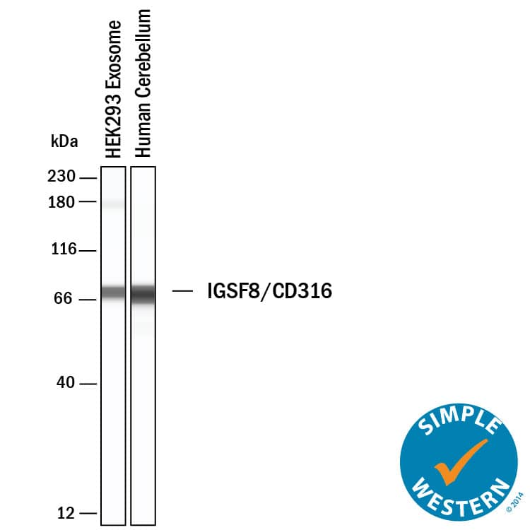
Detection of Human IGSF8/CD316 by Simple WesternTM. Simple Western lane view shows lysates of Exosome Standards (HEK293) (NBP3-11684) and human cerebellum tissue, loaded at 0.5 mg/ml. A specific band was detected for IGSF8/CD316 at approximately 69 kDa (as indicated) using 20 µg/ml of Goat Anti-Human/Mouse/Rat IGSF8/CD316 Antigen Affinity-purified Polyclonal Antibody (Catalog # AF3117) followed by HRP-conjugated Donkey Anti-Goat Secondary Antibody (Catalog # 042-206). This experiment was conducted under reducing conditions and using the 12-230kDa separation system.
 View Larger
Detection of Rat IGSF8/CD316 by Western Blot. Western blot shows lysates of rat brain (hippocampus) tissue. PVDF membrane was probed with 1 µg/mL of Goat Anti-Human/Mouse/Rat IGSF8/CD316 Antigen Affinity-purified Polyclonal Antibody (Catalog # AF3117) followed by HRP-conjugated Anti-Goat IgG Secondary Antibody (Catalog # HAF017). A specific band was detected for IGSF8/CD316 at approximately 70 kDa (as indicated). This experiment was conducted under reducing conditions and using Immunoblot Buffer Group 1.
 View Larger
Detection of IGSF8/CD316 in Neuro‑2A Mouse Cell Line by Flow Cytometry. Neuro-2A mouse neuroblastoma cell line was stained with Goat Anti-Human/Mouse/Rat IGSF8/CD316 Antigen Affinity-purified Polyclonal Antibody (Catalog # AF3117, filled histogram) or isotype control antibody (Catalog # AB-108-C, open histogram), followed by Phycoerythrin-conjugated Anti-Goat IgG Secondary Antibody (Catalog # F0107).
 View Larger
IGSF8/CD316 in Neuro‑2A Mouse Cell Line. IGSF8/CD316 was detected in immersion fixed Neuro-2A mouse neuroblastoma cell line using Goat Anti-Human/Mouse/Rat IGSF8/CD316 Antigen Affinity-purified Polyclonal Antibody (Catalog # AF3117) at 10 µg/mL for 3 hours at room temperature. Cells were stained using the NorthernLights™ 557-conjugated Anti-Goat IgG Secondary Antibody (red; Catalog # NL001) and counterstained with DAPI (blue). Specific staining was localized to cell surfaces. View our protocol for Fluorescent ICC Staining of Cells on Coverslips.
 View Larger
Detection of Human, Mouse, and Rat IGSF8/CD316 by Simple WesternTM. Simple Western lane view shows lysates of human cerebellum tissue, human hippocampus tissue, Neuro-2A mouse neuroblastoma cell line, and rat hippocampus tissue, loaded at 0.2 mg/mL. A specific band was detected for IGSF8/CD316 at approximately 71-80 kDa (as indicated) using 20 µg/mL of Goat Anti-Human/Mouse IGSF8/CD316 Antigen Affinity-purified Polyclonal Antibody (Catalog # AF3117) followed by 1:50 dilution of HRP-conjugated Anti-Goat IgG Secondary Antibody (Catalog # HAF109). This experiment was conducted under reducing conditions and using the 12-230 kDa separation system.
 View Larger
Detection of Human IGSF8/CD316 by Western Blot WGA-HRP identifies a number of EV-specific markers that are present regardless of oncogene status.(A) Matrix depicting samples analyzed during LFQ comparison–Control and Myc cells, as well as Control and Myc EVs. (B) Principle component analysis (PCA) of all four groups analyzed by LFQ. Component 1 (50.4%) and component 2 (15.8%) are graphed. (C) Functional annotation was performed for each gene cluster using DAVID Bioinformatics Resource 6.8 and the highest ranking annotation features for the EV-specific gene cluster are shown. (D) Heatmap of the 50 most upregulated proteins in either RWPE-1 cells or EVs. Proteins are listed in decreasing order of expression with the most highly expressed proteins in EVs on the far left and the most highly expressed proteins in cells on the far right. Averages from all four replicates of each sample type are graphed. Scale indicates intensity, defined as (LFQ Area−Mean LFQ Area)/Standard Deviation. Extracellular proteins with annotated transmembrane domains are bolded and annotated secreted proteins are italicized. (E) Table indicating fold-change of most differentially regulated proteins by LC-MS/MS for RWPE-1 EVs compared to parent cells. (F) Western blot showing the EV-specific marker ITIH4, IGSF8, and MFGE8. Mass spectrometry data is based on two biological and two technical replicates (N=4). Due to limited sample yield, one replicate was performed for the EV western blot. EV, extracellular vesicle; LFQ, label-free quantification.Figure 5—source data 1.Uncropped western blots.Figure 5—source data 2.Mass spectrometry analysis results table.Figure 5—source data 3.List of proteins comparing enriched targets (>2-fold) in Control EVs versus Control cells and Myc EVs versus Myc cells.Uncropped western blots.Mass spectrometry analysis results table.List of proteins comparing enriched targets (>2-fold) in Control EVs versus Control cells and Myc EVs versus Myc cells.Heatmap comparison of biological and technical replicates of RWPE-1 Control/Myc cells and EVs.Biological and technical replicates cluster together based on both oncogene status and compartment for EV or cell surface. Proteins with no area values were assigned an imputed value using Perseus. Heatmap clustering is based off of the Pearson correlation between all replicates on both columns and rows. Heatmap was produced using Morpheus, https://software.broadinstitute.org/morpheus. The first number following the sample name denotes the biological replicated and second number denotes the technical replicate. Image collected and cropped by CiteAb from the following publication (https://pubmed.ncbi.nlm.nih.gov/35257663), licensed under a CC-BY license. Not internally tested by R&D Systems.
Reconstitution Calculator
Preparation and Storage
- 12 months from date of receipt, -20 to -70 °C as supplied.
- 1 month, 2 to 8 °C under sterile conditions after reconstitution.
- 6 months, -20 to -70 °C under sterile conditions after reconstitution.
Background: IGSF8/CD316
IGSF8, also known as PGRL (PG Regulatory-Like Protein), KASP (KAI/CD82 Associated Protein) and EWI-2 (Glu-Trp-Ile motif 2), is a widely expressed transmembrane adhesion protein. It interacts with beta 1 Integrins and various tetraspanins including CD9, CD81 and CD82. IGSF8 contains four extracellular Ig-like domains. IGSF8 over-expression in transformed cells inhibits cell migration and suppresses cancer metastatic potential. Mouse and human IGSF8 share 90% amino acid sequence identity.
Product Datasheets
Citations for Human/Mouse/Rat IGSF8/CD316 Antibody
R&D Systems personnel manually curate a database that contains references using R&D Systems products. The data collected includes not only links to publications in PubMed, but also provides information about sample types, species, and experimental conditions.
11
Citations: Showing 1 - 10
Filter your results:
Filter by:
-
Bacterial meningitis in the early postnatal mouse studied at single-cell resolution
Authors: Wang J, Rattner A, Nathans J
eLife
-
Cell-surface tethered promiscuous biotinylators enable comparative small-scale surface proteomic analysis of human extracellular vesicles and cells
Authors: Lisa L Kirkemo, Susanna K Elledge, Jiuling Yang, James R Byrnes, Jeff E Glasgow, Robert Blelloch et al.
eLife
-
Microscopic clusters feature the composition of biochemical tetraspanin-assemblies and constitute building-blocks of tetraspanin enriched domains
Authors: Schmidt, SC;Massenberg, A;Homsi, Y;Sons, D;Lang, T;
Scientific reports
Species: Human
Sample Types: Whole Cells
Applications: ICC -
MicroRNA-7 regulates melanocortin circuits involved in mammalian energy homeostasis
Authors: MP LaPierre, K Lawler, S Godbersen, IS Farooqi, M Stoffel
Nature Communications, 2022-09-29;13(1):5733.
Species: Mouse
Sample Types: Cell Lysates
Applications: Western Blot -
A conserved sequence in the small intracellular loop of tetraspanins forms an M-shaped inter-helix turn
Authors: N Reppert, T Lang
Scientific Reports, 2022-03-16;12(1):4494.
Species: Human
Sample Types: Cell Lysates
Applications: Western Blot -
EWI‐2 controls nucleocytoplasmic shuttling of EGFR signaling molecules and miRNA sorting in exosomes to inhibit prostate cancer cell metastasis
Authors: Chenying Fu, Qing Zhang, Ani Wang, Songpeng Yang, Yangfu Jiang, Lin Bai et al.
Molecular Oncology
-
Hspa8 and ICAM-1 as damage-induced mediators of &gamma&delta T cell activation
Authors: MD Johnson, MF Otuki, DA Cabrini, R Rudolph, DA Witherden, WL Havran
Journal of leukocyte biology, 2021-04-13;0(0):.
Species: Mouse
Sample Types: Whole Cells
Applications: Flow Cytometry -
Synapse type-specific proteomic dissection identifies IgSF8 as a hippocampal CA3 microcircuit organizer
Authors: N Apóstolo, SN Smukowski, J Vanderlind, G Condomitti, V Rybakin, J Ten Bos, L Trobiani, S Portegies, KM Vennekens, NV Gounko, D Comoletti, KD Wierda, JN Savas, J de Wit
Nat Commun, 2020-10-14;11(1):5171.
Species: Mouse, Rat
Sample Types: Cell Culture Supernates, Whole Tissue
Applications: ICC, IHC -
Transcriptome analysis reveals transmembrane targets on transplantable midbrain dopamine progenitors.
Authors: Bye C, Jonsson M, Bjorklund A, Parish C, Thompson L
Proc Natl Acad Sci U S A, 2015-03-09;112(15):E1946-55.
Species: Mouse, Rat
Sample Types: Tissue Homogenates, Whole Tissue
Applications: Flow Cytometry, IHC -
EWI-2 negatively regulates TGF-beta signaling leading to altered melanoma growth and metastasis.
Authors: Wang H, Sharma C, Knoblich K, Granter S, Hemler M
Cell Res, 2015-02-06;25(3):370-85.
Species: Mouse
Sample Types: Whole Tissue
Applications: IHC -
IgSF8: a developmentally and functionally regulated cell adhesion molecule in olfactory sensory neuron axons and synapses.
Authors: Ray A, Treloar H
Mol Cell Neurosci, 2012-06-09;50(3):238-49.
Species: Mouse
Sample Types: Cell Lysates, Whole Tissue
Applications: IHC-Fr, Immunoprecipitation, Western Blot
FAQs
No product specific FAQs exist for this product, however you may
View all Antibody FAQsReviews for Human/Mouse/Rat IGSF8/CD316 Antibody
Average Rating: 5 (Based on 1 Review)
Have you used Human/Mouse/Rat IGSF8/CD316 Antibody?
Submit a review and receive an Amazon gift card.
$25/€18/£15/$25CAN/¥75 Yuan/¥2500 Yen for a review with an image
$10/€7/£6/$10 CAD/¥70 Yuan/¥1110 Yen for a review without an image
Filter by:

