Human/Mouse/Rat Jak1 Antibody Summary
Pro32-Phe286
Accession # P23458
*Small pack size (-SP) is supplied either lyophilized or as a 0.2 µm filtered solution in PBS.
Applications
Please Note: Optimal dilutions should be determined by each laboratory for each application. General Protocols are available in the Technical Information section on our website.
Scientific Data
 View Larger
Detection of Human and Mouse Jak1 by Western Blot. Western blot shows lysates of Jurkat human acute T cell leukemia cell line, K562 human chronic myelogenous leukemia cell line, A20 mouse B cell lymphoma cell line, and L1.2 mouse pro-B cell line. PVDF membrane was probed with 1 µg/mL of Rat Anti-Human/Mouse/Rat Jak1 Monoclonal Antibody (Catalog # MAB4260) followed by HRP-conjugated Anti-Rat IgG Secondary Antibody (Catalog # HAF005). A specific band was detected for Jak1 at approximately 130 kDa (as indicated). This experiment was conducted under reducing conditions and using Immunoblot Buffer Group 3.
 View Larger
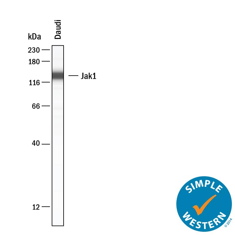
Detection of Human/Mouse/Rat Jak1 by Simple WesternTM. Simple Western lane view shows lysates of Daudi human Burkitt's lymphoma cell line, loaded at 0.2 mg/mL. A specific band was detected for Jak1 at approximately 136 kDa (as indicated) using 10 µg/mL of Rat Anti-Human/Mouse/Rat Jak1 Monoclonal Antibody (Catalog # MAB4260). This experiment was conducted under reducing conditions and using the 12-230 kDa separation system.
Reconstitution Calculator
Preparation and Storage
- 12 months from date of receipt, -20 to -70 °C as supplied.
- 1 month, 2 to 8 °C under sterile conditions after reconstitution.
- 6 months, -20 to -70 °C under sterile conditions after reconstitution.
Background: Jak1
Janus Kinase 1 (Jak1) belongs to a family of protein tyrosine kinases that couple to cytokine receptors and are activated by ligand binding to these receptors. Activation of Jak1 occurs via phosphorylation at two adjacent tyrosine residues, Y1022 and Y1023, within the kinase domain. Jaks activate members of the STAT family of transcription factors by phosphorylating critical tyrosine regulatory sites. Jak1 is required for the activation of STAT1 and STAT2 in response to interferon alpha.
Product Datasheets
Product Specific Notices
This product is sold under license from Millipore Corporation under the following US or foreign patents: 5,821,069; 5,658,791; EP0560890. This product shall not be used to commercially screen drug molecules being developed as JAK1 or JAK2 inhibitors. Any such activity will be outside the scope of the research use only label license.Citations for Human/Mouse/Rat Jak1 Antibody
R&D Systems personnel manually curate a database that contains references using R&D Systems products. The data collected includes not only links to publications in PubMed, but also provides information about sample types, species, and experimental conditions.
2
Citations: Showing 1 - 2
Filter your results:
Filter by:
-
Single-Cell Spatial MIST for Versatile, Scalable Detection of Protein Markers
Authors: Meah, A;Vedarethinam, V;Bronstein, R;Gujarati, N;Jain, T;Mallipattu, SK;Li, Y;Wang, J;
Biosensors
Species: Mouse
Sample Types: Complex Sample Type
Applications: IHC -
3-oxo-C12:2-HSL, quorum sensing molecule from human intestinal microbiota, inhibits pro-inflammatory pathways in immune cells via bitter taste receptors
Authors: G Coquant, D Aguanno, L Brot, C Belloir, J Delugeard, N Roger, HP Pham, L Briand, M Moreau, L de Sordi, V Carrière, JP Grill, S Thenet, P Seksik
Oncogene, 2022-06-08;12(1):9440.
Species: Mouse
Sample Types: Cell Lysates
Applications: Western Blot
FAQs
-
Will the Human/Mouse/Rat Jak1 Antibody, Catalog # MAB4260, detect both phosphorylated and unphosphorylated JAK1?
The Human/Mouse/Rat Jak1 Antibody, Catalog # MAB4260, was raised against an E. coli-derived recombinant human Jak1 (Pro32-Phe286) protein, Accession # P23458. The epitope detected by the antibody is in this region. Theoretically, the antibody would react with both phosphorylated and unphosphorylated JAK1. However, we have not attempted to confirm this experimentally.
Reviews for Human/Mouse/Rat Jak1 Antibody
Average Rating: 5 (Based on 3 Reviews)
Have you used Human/Mouse/Rat Jak1 Antibody?
Submit a review and receive an Amazon gift card.
$25/€18/£15/$25CAN/¥75 Yuan/¥2500 Yen for a review with an image
$10/€7/£6/$10 CAD/¥70 Yuan/¥1110 Yen for a review without an image
Filter by:


