Human/Mouse/Rat RelA/NF kappa B p65 Antibody Summary
Asn456-Ser551
Accession # Q04206
*Small pack size (-SP) is supplied either lyophilized or as a 0.2 µm filtered solution in PBS.
Applications
Please Note: Optimal dilutions should be determined by each laboratory for each application. General Protocols are available in the Technical Information section on our website.
Scientific Data
 View Larger
Detection of Human RelA/NF kappa B p65 by Western Blot. Western Blot shows lysates of HeLa human cervical epithelial carcinoma cell line and K562 human chronic myelogenous leukemia cell line. PVDF membrane was probed with 2 µg/ml of Mouse Anti-Human/Mouse/Rat RelA/NF kappa B p65 Monoclonal Antibody (Catalog # MAB5078) followed by HRP-conjugated Anti-Mouse IgG Secondary Antibody (Catalog # HAF018). A specific band was detected for RelA/NF kappa B p65 at approximately 65 kDa (as indicated). This experiment was conducted under reducing conditions and using Western Blot Buffer Group 1.
 View Larger
Detection of Mouse RelA/NF kappa B p65 by Western Blot. Western Blot shows lysates of BaF3 mouse pro-B cell line. PVDF membrane was probed with 2 µg/ml of Mouse Anti-Human/Mouse/Rat RelA/NF kappa B p65 Monoclonal Antibody (Catalog # MAB5078) followed by HRP-conjugated Anti-Mouse IgG Secondary Antibody (Catalog # HAF018). A specific band was detected for RelA/NF kappa B p65 at approximately 65 kDa (as indicated). This experiment was conducted under reducing conditions and using Western Blot Buffer Group 1.
 View Larger
Detection of Rat RelA/NF kappa B p65 by Western Blot. Western blot shows lysates of C6 rat glioma cell line. PVDF membrane was probed with 2 µg/mL of Mouse Anti-Human/Mouse/Rat RelA/NF kappa B p65 Monoclonal Antibody (Catalog # MAB5078) followed by HRP-conjugated Anti-Mouse IgG Secondary Antibody (Catalog # HAF018). A specific band was detected for RelA/NF kappa B p65 at approximately 65 kDa (as indicated). This experiment was conducted under reducing conditions and using Immunoblot Buffer Group 1.
 View Larger
RelA/NF kappa B p65 in K562 Human Cell Line. RelA/NF kappa B p65 was detected in immersion fixed K562 human chronic myelogenous leukemia cell line using Mouse Anti-Human/Mouse/Rat RelA/NF kappa B p65 Monoclonal Antibody (Catalog # MAB5078) at 10 µg/mL for 3 hours at room temperature. Cells were stained using the NorthernLights™ 557-conjugated Anti-Mouse IgG Secondary Antibody (red; Catalog # NL007) and counterstained with DAPI (blue). View our protocol for Fluorescent ICC Staining of Non-adherent Cells.
 View Larger
Detection of RelA in HeLa Human Cell Line by Flow Cytometry. HeLa human cervical epithelial carcinoma cell line was stained with Mouse Anti-Human/Mouse/Rat RelA/NF kappa B p65 Monoclonal Antibody (Catalog # MAB5078, filled histogram) or isotype control antibody (Catalog # MAB0041, open histogram), followed by Phycoerythrin-conjugated Anti-Mouse IgG Secondary Antibody (Catalog # F0102B). To facilitate intracellular staining, cells were fixed with paraformadehyde and permeabilized with methanol.
 View Larger
Western Blot Shows Human RelA/NF kappa B p65 Specificity by Using Knockout Cell Line. Western blot shows lysates of HeLa human cervical epithelial carcinoma parental cell line and RelA/NF kappa B p65 knockout HeLa cell line (KO). PVDF membrane was probed with 2 µg/mL of Mouse Anti-Human/Mouse/Rat RelA/NF kappa B p65 Monoclonal Antibody (Catalog # MAB5078) followed by HRP-conjugated Anti-Mouse IgG Secondary Antibody (Catalog # HAF007). A specific band was detected for RelA/NF kappa B p65 at approximately 65 kDa (as indicated) in the parental HeLa cell line, but is not detectable in knockout HeLa cell line. GAPDH (Catalog # AF5718) is shown as a loading control. This experiment was conducted under reducing conditions and using Immunoblot Buffer Group 1.
 View Larger
Detection of Human RelA/NFkB p65 by Western Blot Non-classical and intermediate monocytes express high levels of NF-kappa B (p65) and membrane-bound IL-1 alpha.a Western blot analysis of total p65 and GAPDH protein levels in the three monocyte subsets. b Quantification of Western blot data shown in (a): p65 protein level was normalized to GAPDH (loading control) and expressed as a fold change with respect to CL subset. The data represent the means ± SD; n = 3. c Relative levels of phosphorylated-p65 (p-p65), measured by flow cytometry. Each line represents one donor; n = 7. d IL-1 alpha secretion by the three monocyte subsets, measured by Luminex assay. The data represent the means ± SD; n = 3. e–f Relative expression levels of membrane-bound IL-1 alpha (e) and cytoplasmic IL-1 alpha (f), analyzed by flow cytometry. Each line represents one donor; n = 9. *p < 0.05; **p < 0.01; ***p < 0.001; ****p < 0.0001. CL: classical, ITM: intermediate, NC: non-classical, MFI: median fluorescence intensity Image collected and cropped by CiteAb from the following publication (https://www.nature.com/articles/s41419-018-0327-1), licensed under a CC-BY license. Not internally tested by R&D Systems.
 View Larger
Detection of Human RelA/NFkB p65 by Western Blot Tumor‐derived EVs activate the NF kappa B pathway. Primary CD14+ monocytes were incubated with TEVs isolated from conditioned PCI‐1 supernatants. (A) Lysates were generated at different timepoints of incubation and tested for I kappa B and phosphorylated P‐I kappa B. Tubulin was used as a loading control. (B) Primary monocytes were incubated with complete PCI‐1 supernatant (SN), in TEV‐depleted SN (depl.) or with isolated TEV for 120 min, nuclei were isolated and the translocation of NF kappa B‐p65 was tested with an immunoblot. Actin was used as a loading control, and the cytoplasmic fraction (p65/cyto) was used to confirm the specificity of the translocation. (C) Monocytes were incubated as in (B), and the binding capacity of NF kappa B p50 to the DNA was quantified with a NF kappa B binding assay kit. Image collected and cropped by CiteAb from the following publication (https://pubmed.ncbi.nlm.nih.gov/29601673), licensed under a CC-BY license. Not internally tested by R&D Systems.
 View Larger
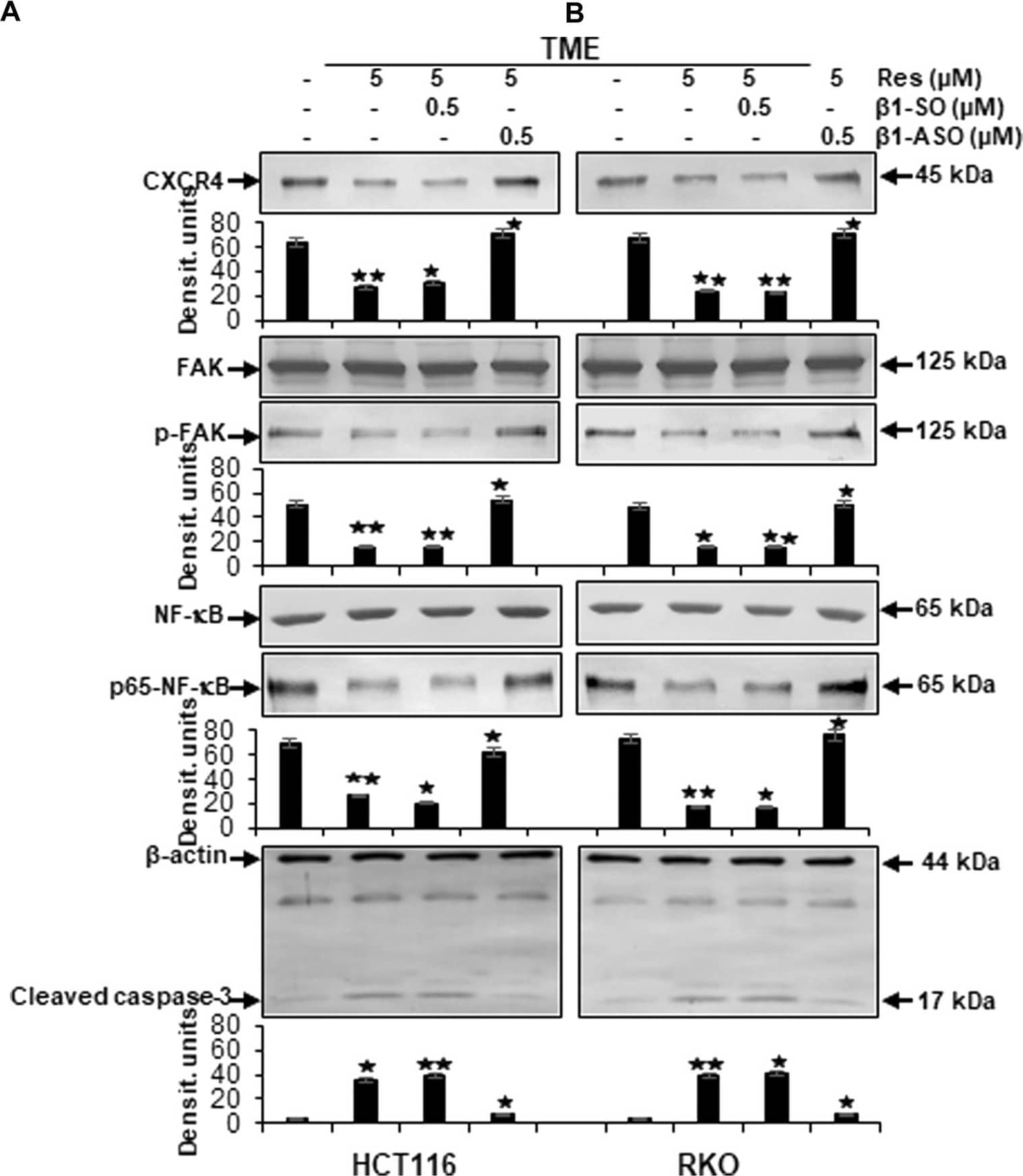
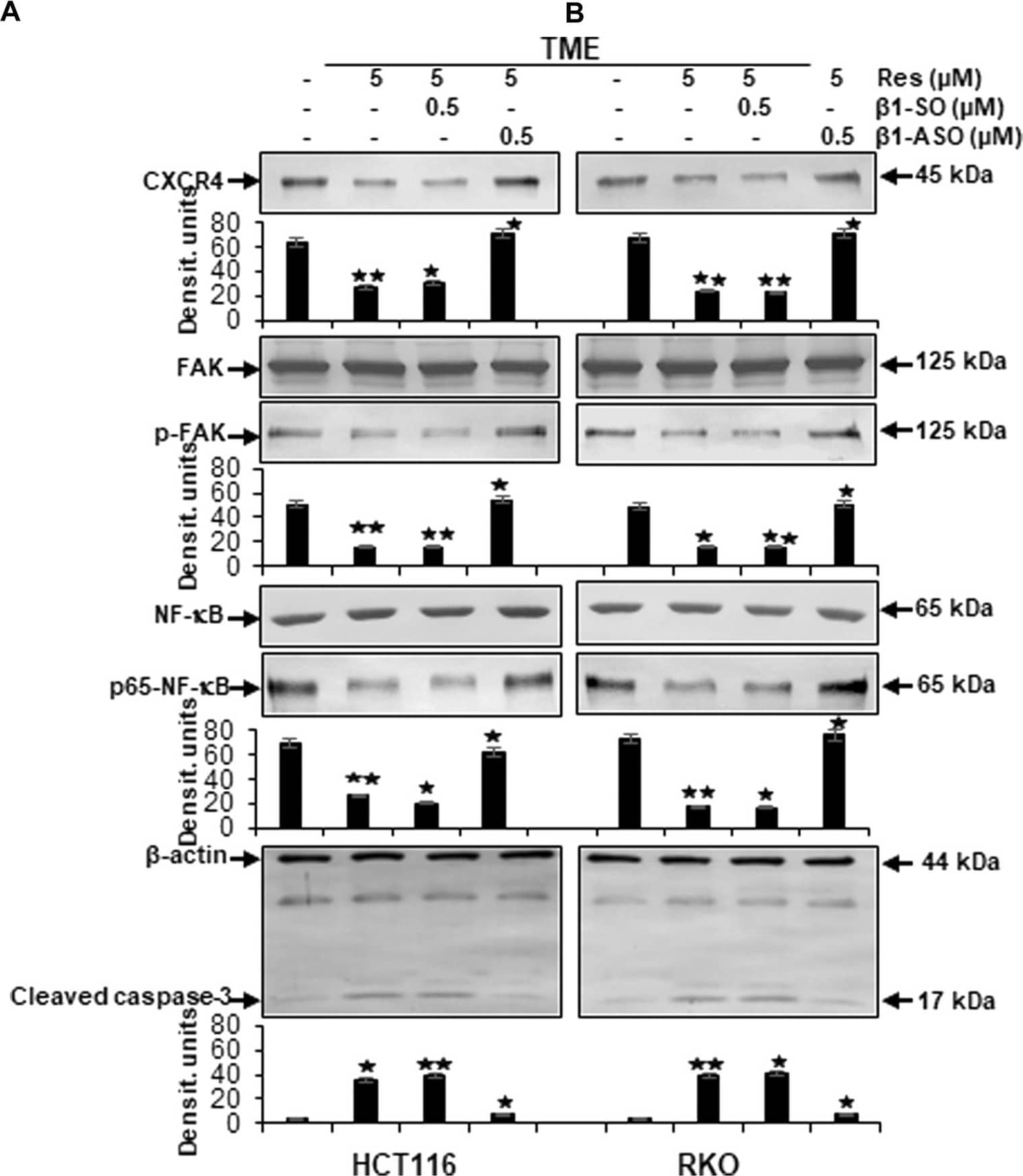
Detection of RelA/NF kappa B p65 by Western Blot Impact of resveratrol or/and ASO against beta 1-integrin on TME-promoted activation of metastasis and apoptosis parameters in CRC cells. HCT116 (A) and RKO (B) derived from 3D-alginate cultures were grown untreated or treated with 5 µM resveratrol alone or in combination with 0.5 µM beta 1-SO or 0.5 µM beta 1-ASO (x-axis) and probed with antibodies against CXCR4, FAK, p-FAK, NF-kappa B, p65-NF-kappa B and cleaved caspase-3. Loading control: beta -actin. Densitometric units complementing Western blot results (y-axis). For densitometric analysis, data were compared to TME control: *p < 0.05 and **p < 0.01. Image collected and cropped by CiteAb from the following open publication (https://pubmed.ncbi.nlm.nih.gov/36120305), licensed under a CC-BY license. Not internally tested by R&D Systems.
Reconstitution Calculator
Preparation and Storage
- 12 months from date of receipt, -20 to -70 °C as supplied.
- 1 month, 2 to 8 °C under sterile conditions after reconstitution.
- 6 months, -20 to -70 °C under sterile conditions after reconstitution.
Background: RelA/NFkB p65
RelA p65 (v-rel reticuloendotheliosis viral oncogene homolog A) is a 65 kDa member of the NF kappa B family of nuclear transcription factors. Dimers of p65 with the p50 subunit are the most common form of the NF kappa B transcription factor, but dimers with it or other family members can also occur. Upon activation, RelA p65 forms an heterotetramer and moves into the nucleus where it binds to specific DNA sequences. An alternatively spliced isoform that lacks amino acids (aa) 222‑231 (p65 delta ) does not bind DNA. Over the sequence used as an immunogen, human RelA p65 shares 96% and 98% aa identity with mouse and rat RelA p65, respectively. This portion includes one of eight potential ser/thr phosphorylation sites, two acetylation sites, and most of the Rel homology domain that interacts with I kappa B inhibitors.
Product Datasheets
Citations for Human/Mouse/Rat RelA/NF kappa B p65 Antibody
R&D Systems personnel manually curate a database that contains references using R&D Systems products. The data collected includes not only links to publications in PubMed, but also provides information about sample types, species, and experimental conditions.
12
Citations: Showing 1 - 10
Filter your results:
Filter by:
-
Inhibition of interleukin-1 signaling enhances elimination of tyrosine kinase inhibitor treated CML stem cells
Blood, 2016-09-12;0(0):.
-
RELA 8-Oxoguanine DNA Glycosylase1 Is an Epigenetic Regulatory Complex Coordinating the Hexosamine Biosynthetic Pathway in RSV Infection
Authors: Xu X, Qiao D, Pan L et al.
Cells
-
Resveratrol Modulates Chemosensitisation to 5-FU via beta1-Integrin/HIF-1alpha Axis in CRC Tumor Microenvironment
Authors: A Brockmuell, S Girisa, AB Kunnumakka, M Shakibaei
International Journal of Molecular Sciences, 2023-03-05;24(5):.
-
Endothelial specific LAT1 ablation normalizes tumor vasculature
Authors: Suehiro, JI;Kimura, T;Fukutomi, T;Naito, H;Kanki, Y;Wada, Y;Kubota, Y;Takakura, N;Sakurai, H;
JCI insight
Species: Human
Sample Types: Cell Lysates, Nuclear Extract
Applications: Western Blot -
Single-Cell Spatial MIST for Versatile, Scalable Detection of Protein Markers
Authors: Meah, A;Vedarethinam, V;Bronstein, R;Gujarati, N;Jain, T;Mallipattu, SK;Li, Y;Wang, J;
Biosensors
Species: Mouse
Sample Types: Complex Sample Type
Applications: IHC -
Calebin A targets the HIF-1?/NF-?B pathway to suppress colorectal cancer cell migration
Authors: Brockmueller, A;Girisa, S;Motallebi, M;Kunnumakkara, AB;Shakibaei, M;
Frontiers in pharmacology
Species: Human
Sample Types: Cell Lysates
Applications: Immunoprecipitation -
Elderberry extract improves molecular markers of endothelial dysfunction linked to atherosclerosis
Authors: Festa J, Hussain A, Hackney A et al.
Food Science & Nutrition
-
beta1-Integrin plays a major role in resveratrol-mediated anti-invasion effects in the CRC microenvironment
Authors: A Brockmuell, AL Mueller, P Shayan, M Shakibaei
Frontiers in Pharmacology, 2022-09-02;13(0):978625.
Species: Human
Sample Types: Whole Cell
Applications: ICC/IF -
Radiotherapy Combined with PD-1 Inhibition Increases NK Cell Cytotoxicity towards Nasopharyngeal Carcinoma Cells
Authors: A Makowska, N Lelabi, C Nothbaum, L Shen, P Busson, TTB Tran, M Eble, U Kontny
Cells, 2021-09-17;10(9):.
Species: Human
Sample Types: Whole Cells
Applications: Flow Cytometry -
Intermittent Hypoxia Activates Duration-Dependent Protective and Injurious Mechanisms in Mouse Lung Endothelial Cells
Authors: P Wohlrab, L Soto-Gonza, T Benesch, MP Winter, IM Lang, K Markstalle, V Tretter, KU Klein
Front Physiol, 2018-12-06;9(0):1754.
Species: Mouse
Sample Types: Cell Lysates
Applications: Western Blot -
Tumor‐derived extracellular vesicles activate primary monocytes
Authors: Kathrin Gärtner, Christina Battke, Judith Dünzkofer, Corinna Hüls, Bettina von Neubeck, Mar‐ kus Kellner et al.
Cancer Medicine
-
The pro-inflammatory phenotype of the human non-classical monocyte subset is attributed to senescence
Authors: SM Ong, E Hadadi, TM Dang, WH Yeap, CT Tan, TP Ng, A Larbi, SC Wong
Cell Death Dis, 2018-02-15;9(3):266.
Species: Human
Sample Types: Cell Lysates
Applications: Western Blot
FAQs
No product specific FAQs exist for this product, however you may
View all Antibody FAQsReviews for Human/Mouse/Rat RelA/NF kappa B p65 Antibody
Average Rating: 5 (Based on 2 Reviews)
Have you used Human/Mouse/Rat RelA/NF kappa B p65 Antibody?
Submit a review and receive an Amazon gift card.
$25/€18/£15/$25CAN/¥75 Yuan/¥2500 Yen for a review with an image
$10/€7/£6/$10 CAD/¥70 Yuan/¥1110 Yen for a review without an image
Filter by:
Paired with AF5078 in ELISA.
1x10^6 MCF7 cells were stained with 2.5 ug NFKBp65 antibody (red) and control antibody (blue). Blocked with 1% BSA, unfixed. Secondary antibody: PE-goat anti-mouse IgG with dilution 1:100.


