Human/Mouse TAB1 Antibody Summary
Ser7-Glu159
Accession # Q15750
Applications
Please Note: Optimal dilutions should be determined by each laboratory for each application. General Protocols are available in the Technical Information section on our website.
Scientific Data
 View Larger
Detection of Human/Mouse TAB1 by Western Blot. Western blot shows lysates of WS-1 human fetal skin fibroblast cell line and C2C12 mouse myoblast cell line. PVDF membrane was probed with 1 µg/mL of Goat Anti-Human/Mouse TAB1 Antigen Affinity-purified Polyclonal Antibody (Catalog # AF3578) followed by HRP-conjugated Anti-Goat IgG Secondary Antibody (Catalog # HAF109). A specific band was detected for TAB1 at approximately 65 kDa (as indicated). This experiment was conducted under reducing conditions and using Immunoblot Buffer Group 2.
 View Larger
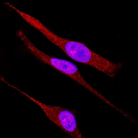
TAB1 in HeLa Human Cell Line. TAB1 was detected in immersion fixed HeLa human cervical epithelial carcinoma cell line using Goat Anti-Human/Mouse TAB1 Antigen Affinity-purified Polyclonal Antibody (Catalog # AF3578) at 15 µg/mL for 3 hours at room temperature. Cells were stained using the NorthernLights™ 557-conjugated Anti-Goat IgG Secondary Antibody (red; Catalog # NL001) and counterstained with DAPI (blue). Specific staining was localized to cytoplasm. View our protocol for Fluorescent ICC Staining of Cells on Coverslips.
Reconstitution Calculator
Preparation and Storage
- 12 months from date of receipt, -20 to -70 °C as supplied.
- 1 month, 2 to 8 °C under sterile conditions after reconstitution.
- 6 months, -20 to -70 °C under sterile conditions after reconstitution.
Background: TAB1
TAK1-binding protein 1 (TAB1), also known as MAP3K7IP1, is an activator of the MAP3K TAK1. The C-terminal 68 amino acids of TAB1 are sufficient for binding and activation of TAK1, while a portion of the N-terminus acts as a dominant-negative inhibitor of TGF-beta, suggesting that TAB1 may function as a signaling intermediate between TGF-beta receptors and TAK1. TAB1 also promotes autophosphorylation and activation of the MAPK p38 alpha.
Product Datasheets
Citations for Human/Mouse TAB1 Antibody
R&D Systems personnel manually curate a database that contains references using R&D Systems products. The data collected includes not only links to publications in PubMed, but also provides information about sample types, species, and experimental conditions.
3
Citations: Showing 1 - 3
Filter your results:
Filter by:
-
BRD4-specific PROTAC inhibits basal-like breast cancer partially through downregulating KLF5 expression
Authors: Kong, Y;Lan, T;Wang, L;Gong, C;Lv, W;Zhang, H;Zhou, C;Sun, X;Liu, W;Huang, H;Weng, X;Cai, C;Peng, W;Zhang, M;Jiang, D;Yang, C;Liu, X;Rao, Y;Chen, C;
Oncogene
Species: Human
Sample Types: Whole Tissue
Applications: Immunohistochemistry -
The Fbw7 tumor suppressor targets KLF5 for ubiquitin-mediated degradation and suppresses breast cell proliferation.
Authors: Zhao D, Zheng HQ, Zhou Z
Cancer Res., 2010-05-18;70(11):4728-38.
Species: Human
Sample Types: Whole Cells
Applications: ICC -
Chlamydia trachomatis antigen induces TLR4-TAB1-mediated inflammation, but not cell death, in maternal decidua cells
Authors: Ortiz B, Driscoll A, Menon R et al.
American journal of reproductive immunology (New York, N.Y. : 1989)
FAQs
No product specific FAQs exist for this product, however you may
View all Antibody FAQsReviews for Human/Mouse TAB1 Antibody
There are currently no reviews for this product. Be the first to review Human/Mouse TAB1 Antibody and earn rewards!
Have you used Human/Mouse TAB1 Antibody?
Submit a review and receive an Amazon gift card.
$25/€18/£15/$25CAN/¥75 Yuan/¥2500 Yen for a review with an image
$10/€7/£6/$10 CAD/¥70 Yuan/¥1110 Yen for a review without an image


