Human/Mouse Wnt-11 Antibody Summary
Leu39-Ala79 and Lys225-Arg297
Accession # Q059Y4
Applications
Please Note: Optimal dilutions should be determined by each laboratory for each application. General Protocols are available in the Technical Information section on our website.
Scientific Data
 View Larger
Wnt‑11 in LNCaP Human Cell Line. Wnt-11 was detected in immersion fixed LNCaP human prostate cancer cell line using Goat Anti-Mouse Wnt-11 Antigen Affinity-purified Polyclonal Antibody (Catalog # AF2647) at 10 µg/mL for 3 hours at room temperature. Cells were stained using the NorthernLights™ 557-conjugated Anti-Goat IgG Secondary Antibody (red; Catalog # NL001) and counterstained with DAPI (blue). Specific staining was localized to cytoplasm. View our protocol for Fluorescent ICC Staining of Cells on Coverslips.
 View Larger
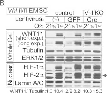
Detection of Mouse Wnt-11 by Western Blot Hypoxia induces expression of WNT11 through VHL.(A,B) Higher basal levels of WNT11 protein in Vhl-deleted cells (lenti-Cre infected Vhlf/f). EMSCs isolated from Vhlf/f mouse were infected with lentivirus carrying either GFP gene (for control) or Cre recombinase (for knockout). Non-infected cells were also used as a control. Immunoblot analysis of control or Vhl KO EMSCs treated with 0.1 mM DMOG (A), and EMSCs exposed to air (21% O2) or hypoxia (1% O2) for 24 hrs (B). Laminin, alpha -tubulin, and lamin A/C were used as loading controls, WNT11 normalized to alpha -Tubulin was shown. (C,D) Inactivation of the Vhl gene results in increased Wnt11 mRNA. Wnt11 and Vegf mRNA levels in liver (C) or duodenum (D) were measured by qPCR in Liver-VhlcKO or duodenum-VhlcKO and control mice (n = 5 per group). Values normalized to Tbp mRNA are expressed relative to tissues from control mice. For panels (C,D), values are mean ± s.e.m. *p < 0.05, **p < 0.01. Image collected and cropped by CiteAb from the following publication (https://www.nature.com/articles/srep21520), licensed under a CC-BY license. Not internally tested by R&D Systems.
 View Larger
Detection of Human Wnt-11 by Western Blot WNT11 is induced by hypoxia or hypoxic mimetics in different cell types.(A) Increased Wnt11 mRNA in EMSC adipocytes (Day 12) after hypoxia-mimetic treatments. EMSC adipocytes were treated with CoCl2 (0.1 mM), DFO (0.1 mM) or DMOG (0.1 mM) for 24 hrs. Values were normalized to Tbp mRNA and are expressed relative to control (n = 3). (B,C) Increased Wnt11 mRNA by hypoxia in EMSC preadipocytes and adipocytes (Day 0–12 after differentiation) (B), and C2C12 myoblast and myocyte (Day 0 and 8 after differentiation) (C). Wnt11 mRNA was assessed by quantitative PCR in cells exposed to air (21% O2) or hypoxia (1% O2) for 24 hrs. (n = 4). Values were normalized to Tbp mRNA and are expressed relative to 21% O2 samples (left panel). (D) Immunoblot analyses of HeLa cells under normal air or hypoxia for 24 hrs. (E,F) Induction of Wnt11 by increasing concentrations of DMOG in MDA-MB-231 cells (E) and 4T1 cells (F). (G) EMSCs treated with 0.1 mM DMOG for the indicated times. Wnt11 and Vegf mRNA expression was measured by qPCR and normalized to Tbp mRNA (n = 4). (H) WNT11 protein levels after DMOG treatment normalized to alpha -Tubulin (upper panel; n = 4). Representative immunoblots of EMSCs treated with 0.1 mM DMOG for the indicated times (Lower panel). (I) Protein expression in MDA-MB-231 cells treated with 0.1 mM DMOG. (J) Induction of Wnt11 promoter activity by hypoxia or hypoxia mimetics. pGL3-Wnt11 promoter plasmid was transfected into C2C12 cells. Cells were incubated with DMOG (left panel, n = 4) or under 21% O2 or 1% O2 (right panel, n = 8) for 24 hrs. For panels (A–C,G,H,J), values are mean ± s.e.m. *p < 0.05, **p < 0.01. For panels of immunoblotting, laminin, alpha -tubulin, and ERK were used as loading controls, WNT11 normalized to alpha -Tubulin was shown. Image collected and cropped by CiteAb from the following publication (https://www.nature.com/articles/srep21520), licensed under a CC-BY license. Not internally tested by R&D Systems.
 View Larger
Detection of Mouse Wnt-11 by Western Blot WNT11 regulates MMPs activities.(A–C) (Top panels): Serum-free medium was conditioned for 24 hrs by the indicated cells, concentrated 20-fold and assayed by gelatin zymography. Gelatinolytic activity is indicated by clear zones against a dark background of stained substrate. (Bottom): Whole cell extracts were immunoblotted with indicated antibodies. (A) Overexpression of Wnt11 in EMSC or BT473 cells enhances activity of MMP-9 and MMP-2. (B) Impaired activity of MMP-9 and MMP-2 in MDA-MB-231 cells (left) or EMSCs (right) stably expressing Wnt11 shRNAs and treated with DMOG. (C) WNT11 is required for MMP-9 and MMP-2 activity in MDA-MB-231 cells (left) or EMSCs (right) under normoxic and hypoxic culture conditions. (D) WNT11 regulates MMP2 protein in media. (Top): conditioned media from indicated cells and treatments. (Bottom): whole cell lysates were immunoblotted with indicated antibodies. (E) Recombinant WNT11 induces both MMP-2 protein and MMP-2 activity in media. (Top panels): Gelatin zymography and immunoblot of serum-free medium conditioned for the indicated times after recombinant WNT11 (r-WNT11) treatment. (Bottom): Whole cell lysates were immunoblotted with indicated antibodies. (F) MMP-2 inhibitor attenuated induced migration by WNT11. MDA-MB-231 cells infected with lentiviruses for stable expression of Wnt11 or GFP (n = 4) were incubated with either vehicle or 1 μM of ARP100. Media in the lower compartment had same concentration of DMSO or inhibitor. Values are mean ± s.e.m. *p < 0.05, **p < 0.01. For panels (A–D), HIF-1 alpha and HIF-2 alpha were shown as a marker of hypoxia, WNT11 normalized to alpha -Tubulin was shown. Image collected and cropped by CiteAb from the following publication (https://www.nature.com/articles/srep21520), licensed under a CC-BY license. Not internally tested by R&D Systems.
 View Larger
Detection of Human Wnt-11 by Western Blot Induced WNT11 expression with tumor hypoxia and WNT11 regulates tumor growth.Antiangiogenic therapy induces WNT11 expression in the orthotopic malignant glioma model. Athymic mice implanted with U87 delta EGFR cells were administered either bevacizumab (6 mg/kg) or vehicle three times per week for 4 weeks. (A) Increased Wnt11 mRNA in xenografts from mice treated with bevacizumab. Values were normalized to HPRT mRNA and are expressed relative to control (n = 10 per group). Values are mean ± s.e.m. *p < 0.05, **p < 0.01. (B) Bevacizumab increased expression of HIF-1 alpha and HIF-2 alpha and WNT11. First 10 lanes are control tumors, and the last 10 lanes are tumors from bevacizumab-treated animals. Lysates from whole tissue and nuclei are indicated. alpha -Tubulin, actin and lamin A/C are loading controls. Image collected and cropped by CiteAb from the following publication (https://www.nature.com/articles/srep21520), licensed under a CC-BY license. Not internally tested by R&D Systems.
 View Larger
Detection of Mouse Wnt-11 by Western Blot HIF-1 alpha is the predominant transcriptional regulator of WNT11 expression during hypoxia.(A,C–E) EMSCs isolated from the indicated mouse genotypes were infected with lentivirus expressing GFP or Cre recombinase. Non-infected cells and GFP infected cells served as controls. Immunoblot analyses of EMSCs derived from the indicated genotypes treated with 0.1 mM DMOG for 24 hrs. (A) Attenuated WNT11 expression in Hif-1 alpha KO EMSCs (lenti-Cre infected Hif-1af/f). (B) HIF-1 alpha regulates WNT11 expression during hypoxia. Impaired WNT11 expression in MDA-MB-231 cells stably expressing HIF-1 alpha shRNAs with hypoxia. Cells were exposed to air (21% O2) or hypoxia (1% O2) for 24 hrs (left panel). Overexpression of HIF-1 alpha in MDA-MB-231 cells enhances WNT11 expression (right panel). (C) Markedly increased WNT11 levels in Hif-1 alpha overexpressing EMSCs (lenti-Cre infected Hif-1 alpha -Tgfl-Stop). (D) Elevated HIF-1 alpha and WNT11 protein after DMOG treatment in HIF-2 alpha KO cells (lenti-Cre infectied Hif-2 alpha fl/fl). (E) Little effect of HIF-2 alpha overexpression on WNT11 protein expression in EMSC (lenti-Cre infectied Hif-2 alpha -Tgfl-Stop). (F) HIF-1 alpha binds to WNT11 and VEGF promoters as assessed by ChIP analyses (n = 3 each condition). (G) WNT11 expression was suppressed in cells with a beta -catenin deficiency. EMSCs stably expressing shRNAs against beta -catenin or scrambled control were treated with 0.1 mM DMOG for 24 hrs and analyzed by immunoblotting. (H) Co-transfection with expression vectors for beta -catenin, Hif-1 alpha and ARNT stimulates further induction of Wnt11 promoter activity. Constructs encoding Hif-1 alpha, Hif-2 alpha, ARNT, beta -catenin and Wnt11 promoter-luciferase reporter, were transiently transfected into HEK293T cells. Cells were harvested and luciferase activities were measured 48 hrs after transfection (n = 3). For panels (A–E,G), laminin, alpha -tubulin, ERK1/2 and lamin A/C were used as loading controls, WNT11 normalized to alpha -Tubulin was shown under the bots. For panels (F,H), values are mean ± s.e.m. *p < 0.05, **p < 0.01 Image collected and cropped by CiteAb from the following publication (https://www.nature.com/articles/srep21520), licensed under a CC-BY license. Not internally tested by R&D Systems.
 View Larger
Detection of Mouse Wnt-11 by Western Blot Hypoxia induces expression of WNT11 through VHL.(A,B) Higher basal levels of WNT11 protein in Vhl-deleted cells (lenti-Cre infected Vhlf/f). EMSCs isolated from Vhlf/f mouse were infected with lentivirus carrying either GFP gene (for control) or Cre recombinase (for knockout). Non-infected cells were also used as a control. Immunoblot analysis of control or Vhl KO EMSCs treated with 0.1 mM DMOG (A), and EMSCs exposed to air (21% O2) or hypoxia (1% O2) for 24 hrs (B). Laminin, alpha -tubulin, and lamin A/C were used as loading controls, WNT11 normalized to alpha -Tubulin was shown. (C,D) Inactivation of the Vhl gene results in increased Wnt11 mRNA. Wnt11 and Vegf mRNA levels in liver (C) or duodenum (D) were measured by qPCR in Liver-VhlcKO or duodenum-VhlcKO and control mice (n = 5 per group). Values normalized to Tbp mRNA are expressed relative to tissues from control mice. For panels (C,D), values are mean ± s.e.m. *p < 0.05, **p < 0.01. Image collected and cropped by CiteAb from the following open publication (https://pubmed.ncbi.nlm.nih.gov/26861754), licensed under a CC-BY license. Not internally tested by R&D Systems.
Reconstitution Calculator
Preparation and Storage
- 12 months from date of receipt, -20 to -70 °C as supplied.
- 1 month, 2 to 8 °C under sterile conditions after reconstitution.
- 6 months, -20 to -70 °C under sterile conditions after reconstitution.
Background: Wnt-11
Wnt-11 belongs to the Wnt family of secreted, highly conserved, cell signaling molecules that play important roles in vertebrate development. Mouse Wnt-11 shares 97% amino acid sequence identity with its human and rat homolog.
Product Datasheets
Citations for Human/Mouse Wnt-11 Antibody
R&D Systems personnel manually curate a database that contains references using R&D Systems products. The data collected includes not only links to publications in PubMed, but also provides information about sample types, species, and experimental conditions.
4
Citations: Showing 1 - 4
Filter your results:
Filter by:
-
Wnt-11 as a Potential Prognostic Biomarker and Therapeutic Target in Colorectal Cancer
Authors: Irantzu Gorroño-Etxebarria, Urko Aguirre, Saray Sanchez, Nerea González, Antonio Escobar, Ignacio Zabalza et al.
Cancers (Basel)
-
Wnt-11 promotes neuroendocrine-like differentiation, survival and migration of prostate cancer cells
Authors: Pinar Uysal-Onganer, Yoshiaki Kawano, Mercedes Caro, Marjorie M Walker, Soraya Diez, R Siobhan Darrington et al.
Molecular Cancer
-
The noncanonical WNT pathway is operative in idiopathic pulmonary arterial hypertension.
Authors: Laumanns IP, Fink L, Wilhelm J, Wolff JC, Mitnacht-Kraus R, Graef-Hoechst S, Stein MM, Bohle RM, Klepetko W, Hoda MA, Schermuly RT, Grimminger F, Seeger W, Voswinckel R
Am. J. Respir. Cell Mol. Biol., 2008-11-21;40(6):683-91.
Species: Human
Sample Types: Whole Tissue
Applications: IHC-P -
Induction of WNT11 by hypoxia and hypoxia-inducible factor-1alpha regulates cell proliferation, migration and invasion.
Authors: Mori H, Yao Y, Learman BS et al.
Sci Rep.
FAQs
No product specific FAQs exist for this product, however you may
View all Antibody FAQsReviews for Human/Mouse Wnt-11 Antibody
There are currently no reviews for this product. Be the first to review Human/Mouse Wnt-11 Antibody and earn rewards!
Have you used Human/Mouse Wnt-11 Antibody?
Submit a review and receive an Amazon gift card.
$25/€18/£15/$25CAN/¥75 Yuan/¥2500 Yen for a review with an image
$10/€7/£6/$10 CAD/¥70 Yuan/¥1110 Yen for a review without an image


