Human MVK Antibody
Human MVK Antibody Summary
Met1-Lys396
Accession # Q03426
Applications
Please Note: Optimal dilutions should be determined by each laboratory for each application. General Protocols are available in the Technical Information section on our website.
Scientific Data
 View Larger
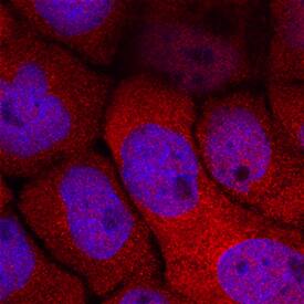
MVK in A431 Human Cell Line. MVK was detected in immersion fixed A431 human epithelial carcinoma cell line using Rabbit Anti-Human MVK Antigen Affinity-purified Polyclonal Antibody (Catalog # AF8516) at 2 µg/mL for 3 hours at room temperature. Cells were stained using the NorthernLights™ 557-conjugated Anti-Rabbit IgG Secondary Antibody (red; Catalog # NL004) and counterstained with DAPI (blue). Specific staining was localized to cytoplasm. View our protocol for Fluorescent ICC Staining of Cells on Coverslips.
 View Larger
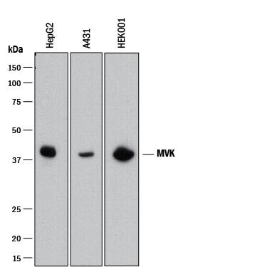
Detection of Human MVK by Western Blot. Western blot shows lysates of HepG2 human hepatocellular carcinoma cell line, A431 human epithelial carcinoma cell line, and HEK001 human epidermal keratinocyte cell line. PVDF membrane was probed with 0.1 µg/mL of Rabbit Anti-Human MVK Antigen Affinity-purified Polyclonal Antibody (Catalog # AF8516) followed by HRP-conjugated Anti-Rabbit IgG Secondary Antibody (Catalog # HAF008). A specific band was detected for MVK at approximately 42 kDa (as indicated). This experiment was conducted under reducing conditions and using Immunoblot Buffer Group 1.
 View Larger
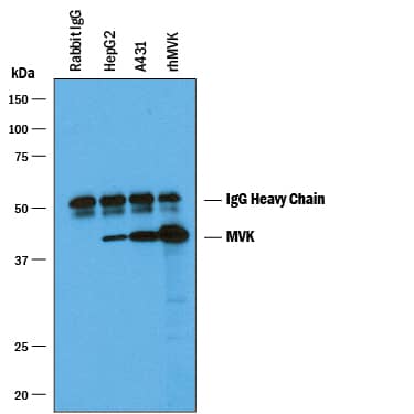
Immunoprecipitation of Human MVK. MVK was immunoprecipitated from 400 µg of HepG2 human hepatocellular carcinoma cell line and A431 human epithelial carcinoma cell line lysates using 4 µg of Rabbit Anti-Human MVK Antigen Affinity-purified Polyclonal Antibody (Catalog # AF8516) coated on 4 wells of a 96 well plate (Corning Costar EIA/RIA). HepG2 lysates, A431 lysates, Rabbit IgG control buffer, or recombinant human MVK were added to the wells and incubated for 2 hours at room temperature. Immunoprecipitated MVK was detected by Western blot under reducing conditions using 0.1 µg/mL Rabbit Anti-Human MVK Antigen Affinity-purified Polyclonal Antibody (Catalog # AF8516) and Immunoblot Buffer Group 1. View our recommended buffer recipes for Immunoprecipitation.
 View Larger
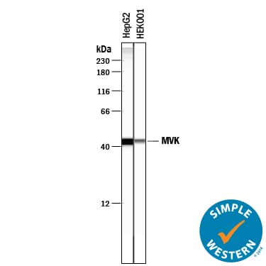
Detection of Human MVK by Simple WesternTM. Simple Western lane view shows lysates of HepG2 human hepatocellular carcinoma cell line and HEK001 human epidermal keratinocyte cell line, loaded at 0.2 mg/mL. A specific band was detected for MVK at approximately 43 kDa (as indicated) using 1 µg/mL of Rabbit Anti-Human MVK Antigen Affinity-purified Polyclonal Antibody (Catalog # AF8516). This experiment was conducted under reducing conditions and using the 12-230 kDa separation system.
Preparation and Storage
- 12 months from date of receipt, -20 to -70 °C as supplied.
- 1 month, 2 to 8 °C under sterile conditions after reconstitution.
- 6 months, -20 to -70 °C under sterile conditions after reconstitution.
Background: MVK
MVK (Mevalonate kinase) is a 42 kDa cytoplasmic protein that belongs to the GHMP kinase family, Mevalonate kinase subfamily of proteins. Mevalonate kinase catalyzes the ATP-dependent phosphorylation of mevalonic acid to form mevalonate 5-phosphate. Defects in mevalonate kinase can cause mevalonic aciduria (MEVA). It is an accumulation of mevalonic acid which causes a variety of symptoms such as psychomotor retardation, dysmorphic features, cataracts, hepatosplenomegaly, lymphadenopathy, anemia, hypotonia, myopathy and ataxia.Over aa 1-396, human MVK shares 81% aa identity with mouse MVK.
Product Datasheets
FAQs
No product specific FAQs exist for this product, however you may
View all Antibody FAQsReviews for Human MVK Antibody
There are currently no reviews for this product. Be the first to review Human MVK Antibody and earn rewards!
Have you used Human MVK Antibody?
Submit a review and receive an Amazon gift card.
$25/€18/£15/$25CAN/¥75 Yuan/¥2500 Yen for a review with an image
$10/€7/£6/$10 CAD/¥70 Yuan/¥1110 Yen for a review without an image

