Human Neudesin Antibody Summary
Gly32-Phe172
Accession # Q9UMX5
Applications
Please Note: Optimal dilutions should be determined by each laboratory for each application. General Protocols are available in the Technical Information section on our website.
Scientific Data
 View Larger
Detection of Human Neudesin by Western Blot. Western blot shows lysates of MCF-7 human breast cancer cell line, IMR-32 human neuroblastoma cell line, and HEK293 human embryonic kidney cell line. PVDF membrane was probed with 2 µg/mL of Mouse Anti-Human Neudesin Monoclonal Antibody (Catalog # MAB6714) followed by HRP-conjugated Anti-Mouse IgG Secondary Antibody (Catalog # HAF007). A specific band was detected for Neudesin at approximately 20 kDa (as indicated). This experiment was conducted under reducing conditions and using Immunoblot Buffer Group 1.
 View Larger
Neudesin in Human Brain. Neudesin was detected in immersion fixed paraffin-embedded sections of human brain (medulla) using Mouse Anti-Human Neudesin Monoclonal Antibody (Catalog # MAB6714) at 15 µg/mL overnight at 4 °C. Before incubation with the primary antibody, tissue was subjected to heat-induced epitope retrieval using Antigen Retrieval Reagent-Basic (Catalog # CTS013). Tissue was stained using the Anti-Mouse HRP-DAB Cell & Tissue Staining Kit (brown; Catalog # CTS002) and counterstained with hematoxylin (blue). Specific staining was localized to cytoplasm of neurons. View our protocol for Chromogenic IHC Staining of Paraffin-embedded Tissue Sections.
 View Larger
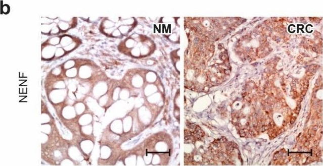
Detection of Neudesin by Immunohistochemistry Characterization of NENF expression and release in colorectal cancer patients and DLD-1 and HT-29 cell lines. NENF expression at gene (a) and protein (b,c) levels in NM (n = 10) and colorectal cancer (CRC) tissues (n = 20). Original magnification, 20×; scale bar, 20 µm. The columns represent the mean ± SEM relative to PPIA. The Mann–Whitney test was used to compare NM vs. CRC results. The differences are statistically non-significant. Serum NENF concentration in colorectal cancer patients (n = 41) compared to the control group (n = 15) (d). The Mann–Whitney test was used to compare serum NENF concentration in C vs. CRC results. Statistical significance: * p ≤ 0.05. NENF expression after treatment with 1 µM of P4 in DLD-1 cell line (e) and HT-29 cell line (f) (n = 3 independent experiments). The Mann–Whitney test was used to compare NENF expression in C vs. CRC results in DLD-1 and HT-29 cells. The differences are statistically non-significant. NENF concentration in the medium after treatment with 1 µM of P4 in DLD-1 cell line (g) (n = 6) and HT-29 cell line (h) (n = 6). The Mann–Whitney test was used to compare NENF concentration in the medium C vs. P4 results of DLD-1 cells, statistical significance: ** p ≤ 0.01. Double staining for PGRMC1 and NENF in NM tissues (n = 10) and colorectal cancer (n = 20) (i). PGRMC1-positive cells are in red, NENF-positive cells are in green, nucleus localization in cells is in blue, and PGRMC1 and NENF merged staining is in orange, scale bar, 20 μm. Abbreviations: C, control/non-treated group; CRC, colorectal cancer; NENF, neudesin; NM, normal mucosa; P4, progesterone; PGRMC1, progesterone receptor membrane component 1; PPIA, peptidylprolyl isomerase A; SD, standard deviation; SEM, standard error of the mean; and vs., versus. Image collected and cropped by CiteAb from the following open publication (https://pubmed.ncbi.nlm.nih.gov/37894441), licensed under a CC-BY license. Not internally tested by R&D Systems.
Reconstitution Calculator
Preparation and Storage
- 12 months from date of receipt, -20 to -70 °C as supplied.
- 1 month, 2 to 8 °C under sterile conditions after reconstitution.
- 6 months, -20 to -70 °C under sterile conditions after reconstitution.
Background: Neudesin
Neudesin (Neuron-derived neurotrophic secreted protein; also CIR2 and SPUF) is a secreted, 20-21 kDa member of the MAPR (membrane-associated progesterone receptor) subfamily, cytochrome b5 family of molecules. It is expressed by CNS neuronal progenitors and neurons, plus preadipocytes in white adipose tissue. Regarding activity, it promotes neuronal differentiation with limited proliferation and serves as a neuron survival factor. By contrast, it inhibits both astrocyte and adipocyte differentiation. Mature human Neudesin is 141 amino acids (aa) in length (aa 32-172). It contains one cytochrome b5-like heme-binding domain (aa 44-129) and an acetylated lysine residue at Lys136. The heme-binding domain does bind heme, and this accounts for 5-6 kDa of its circulating molecular weight. The presence of a heme moiety is necessary for activity. Mature human Neudesin shares 97% aa identity with mature mouse Neudesin
Product Datasheets
Citation for Human Neudesin Antibody
R&D Systems personnel manually curate a database that contains references using R&D Systems products. The data collected includes not only links to publications in PubMed, but also provides information about sample types, species, and experimental conditions.
1 Citation: Showing 1 - 1
-
New Insights on the Progesterone (P4) and PGRMC1/NENF Complex Interactions in Colorectal Cancer Progression
Authors: Kami?ska, J;Koper-Lenkiewicz, OM;Ponikwicka-Tyszko, D;Lebiedzi?ska, W;Palak, E;Sztachelska, M;Bernaczyk, P;Dorf, J;Guzi?ska-Ustymowicz, K;Zar?ba, K;Wo?czy?ski, S;Rahman, NA;Dymicka-Piekarska, V;
Cancers
Species: Human
Sample Types: Whole Cells, Whole Tissue
Applications: ICC, IHC
FAQs
No product specific FAQs exist for this product, however you may
View all Antibody FAQsReviews for Human Neudesin Antibody
There are currently no reviews for this product. Be the first to review Human Neudesin Antibody and earn rewards!
Have you used Human Neudesin Antibody?
Submit a review and receive an Amazon gift card.
$25/€18/£15/$25CAN/¥75 Yuan/¥2500 Yen for a review with an image
$10/€7/£6/$10 CAD/¥70 Yuan/¥1110 Yen for a review without an image
