Human Oct-4A Antibody Summary
Met1-Glu135
Accession # NP_002692
*Small pack size (-SP) is supplied either lyophilized or as a 0.2 µm filtered solution in PBS.
Applications
Please Note: Optimal dilutions should be determined by each laboratory for each application. General Protocols are available in the Technical Information section on our website.
Scientific Data
 View Larger
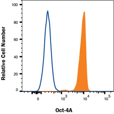
Detection of Oct‑4A in NTERA-2 cells by Flow Cytometry NTERA-2 cells were stained with Mouse Anti-Human Oct‑4A Monoclonal Antibody (Catalog # MAB17591, filled histogram) or isotype control antibody (Catalog # MAB003, open histogram) followed by Allophycocyanin-conjugated Anti-Mouse IgG Secondary Antibody (Catalog # F0101B). To facilitate intracellular staining, cells were fixed and permeabilized with FlowX FoxP3 Fixation & Permeabilization Buffer Kit (Catalog # FC012). View our protocol for Staining Intracellular Molecules.
 View Larger
Detection of Human Oct‑4A by Western Blot. Western blot shows lysates of HeLa human cervical epithelial carcinoma cell line and NTera-2 human testicular embryonic carcinoma cell line. PVDF Membrane was probed with 0.5 µg/mL of Mouse Anti-Human Oct-4A Monoclonal Antibody (Catalog # MAB17591) followed by HRP-conjugated Anti-Mouse IgG Secondary Antibody (HAF007). A specific band was detected for Oct-4A at approximately 50 kDa (as indicated). This experiment was conducted under reducing conditions and using Immunoblot Buffer Group 1.
 View Larger
Oct-4A and E-Cadherin in BG01V Human Stem Cells. Oct-4A and E-Cadherin were detected in human BG01V embryonic stem cells grown on irradiated MEF cells using 10 µg/mL Human Oct-4A Monoclonal Antibody (Catalog # MAB17591) and 10 µg/mL Human E-Cadherin Affinity-purified Polyclonal Antibody (AF648). Cells were incubated with primary antibodies for 3 hours at room temperature. Cells were stained for Oct-4A using the Northern-Lights™ 493-conjugated Anti-Mouse IgG Secondary Antibody (green; NL009), and stained for E-Cadherin using the Northern-Lights™ 557-conjugated Anti-Goat IgG Secondary Antibody (red; NL001). Specific staining of Oct-4A was localized to nuclei. View our protocol for Fluorescent ICC Staining of Cells on Coverslips.
 View Larger
Detection of Oct‑4A in BG01V Human Stem Cells by Flow Cytometry. BG01V human embryonic stem cells were stained with Human Oct-4A Monoclonal Antibody (Catalog # MAB17591, filled histogram) or isotype control antibody (MAB003, open histogram), followed by Allophycocyanin-conjugated Anti-Mouse IgG F(ab')2Secondary Antibody (F0101B). To facilitate intracellular staining, cells were fixed with paraformaldehyde and permeabilized with saponin.
 View Larger
Detection of Oct‑4A in iPSCs by Flow Cytometry. iPSCs were stained with Mouse Anti-Human Oct‑4A Monoclonal Antibody (Catalog # MAB17591, filled histogram) or isotype control antibody (Catalog # MAB003, open histogram), followed by Allophycocyanin-conjugated Anti-Mouse IgG Secondary Antibody (Catalog # F0101B). To facilitate intracellular staining, cells were fixed with Flow Cytometry Fixation Buffer (Catalog # FC004) and permeabilized with Flow Cytometry Permeabilization/Wash Buffer I (Catalog # FC005). View our protocol for Staining Intracellular Molecules.
Reconstitution Calculator
Preparation and Storage
- 12 months from date of receipt, -20 to -70 °C as supplied.
- 1 month, 2 to 8 °C under sterile conditions after reconstitution.
- 6 months, -20 to -70 °C under sterile conditions after reconstitution.
Background: Oct-4A
Oct-3/4, alternately Oct-3 or Oct-4, is POU5F1 (POU domain containing, class 5, transcription factor 1), a 360 amino acid (aa) transcription factor that is expressed in totipotent embryonic stem and germ cells. The human Oct-4, Oct-3/4 or POU5F1 gene can be transcribed into at least three transcripts (Oct-4A, Oct-4B, and Oct-4B1) and generates four protein isoforms by alternative splicing and alternative translation initiation. Oct-4A expression is restricted to embryonic stem (ES) and embryonic carcinoma (EC) cells and is believed to be the transcription factor responsible for the pluripotency properties of embryonic stem (ES) cells. In contrast, Oct-4B/4B1 can be detected in various nonpluripotent cell types and cannot sustain ES cell pluripotency and self-renewal.
Product Datasheets
Citations for Human Oct-4A Antibody
R&D Systems personnel manually curate a database that contains references using R&D Systems products. The data collected includes not only links to publications in PubMed, but also provides information about sample types, species, and experimental conditions.
8
Citations: Showing 1 - 8
Filter your results:
Filter by:
-
ALS-associated KIF5A mutations abolish autoinhibition resulting in a toxic gain of function
Authors: DM Baron, AR Fenton, S Saez-Atien, A Giampetruz, A Sreeram, Shankarach, PJ Keagle, VR Doocy, NJ Smith, EW Danielson, M Andresano, MC McCormack, J Garcia, V Bercier, L Van Den Bo, JR Brent, C Fallini, BJ Traynor, ELF Holzbaur, JE Landers
Cell Reports, 2022-04-05;39(1):110598.
-
Loss of function of the ALS-associated NEK1 kinase disrupts microtubule homeostasis and nuclear import
Authors: Mann, JR;McKenna, ED;Mawrie, D;Papakis, V;Alessandrini, F;Anderson, EN;Mayers, R;Ball, HE;Kaspi, E;Lubinski, K;Baron, DM;Tellez, L;Landers, JE;Pandey, UB;Kiskinis, E;
Science advances
Species: Human
Sample Types: Whole Cells
Applications: ICC -
Expression of ALS-PFN1 impairs vesicular degradation in iPSC-derived microglia
Authors: Funes, S;Gadd, DH;Mosqueda, M;Zhong, J;Jung, J;Shankaracharya, ;Unger, M;Cameron, D;Dawes, P;Keagle, PJ;McDonough, JA;Boopathy, S;Sena-Esteves, M;Lutz, C;Skarnes, WC;Lim, ET;Schafer, DP;Massi, F;Landers, JE;Bosco, DA;
bioRxiv : the preprint server for biology
Species: Human
Sample Types: Whole Cells
Applications: ICC -
Interactions between ALS-linked FUS and nucleoporins are associated with defects in the nucleocytoplasmic transport pathway
Authors: YC Lin, MS Kumar, N Ramesh, EN Anderson, AT Nguyen, B Kim, S Cheung, JA McDonough, WC Skarnes, R Lopez-Gonz, JE Landers, NL Fawzi, IRA Mackenzie, EB Lee, JA Nickerson, D Grunwald, UB Pandey, DA Bosco
Nature Neuroscience, 2021-05-31;0(0):.
Species: Human
Sample Types: Whole Cells
Applications: ICC -
Human mid-trimester amniotic fluid (stem) cells lack expression of the pluripotency marker OCT4A
Authors: F Vlahova, KE Hawkins, AM Ranzoni, KL Hau, R Sagar, P Coppi, AL David, J Adjaye, PV Guillot
Sci Rep, 2019-05-31;9(1):8126.
Species: Human
Sample Types: Cell Lysates, Whole Cells
Applications: Flow Cytometry, Western Blot -
Genome-Wide Transcriptome and Binding Sites Analyses Identify Early FOX Expressions for Enhancing Cardiomyogenesis Efficiency of hESC Cultures
Sci Rep, 2016-08-09;6(0):31068.
Species: Human
Sample Types: Whole Cells
Applications: Flow Cytometry -
Effect of culture medium on propagation and phenotype of corneal stroma–derived stem cells
Authors: Laura E. Sidney, Matthew J. Branch, Harminder S. Dua, Andrew Hopkinson
Cytotherapy
-
Derivation and High Engraftment of Patient-Specific Cardiomyocyte Sheet Using Induced Pluripotent Stem Cells Generated From Adult Cardiac Fibroblast
Authors: Liying Zhang, Jing Guo, Pengyuan Zhang, Qiang Xiong, Steven C. Wu, Lily Xia et al.
Circulation: Heart Failure
FAQs
No product specific FAQs exist for this product, however you may
View all Antibody FAQsReviews for Human Oct-4A Antibody
Average Rating: 4 (Based on 2 Reviews)
Have you used Human Oct-4A Antibody?
Submit a review and receive an Amazon gift card.
$25/€18/£15/$25CAN/¥75 Yuan/¥2500 Yen for a review with an image
$10/€7/£6/$10 CAD/¥70 Yuan/¥1110 Yen for a review without an image
Filter by:


