Human Plexin D1 Antibody Summary
Leu47-Ala1271
Accession # Q9Y4D7
Applications
Please Note: Optimal dilutions should be determined by each laboratory for each application. General Protocols are available in the Technical Information section on our website.
Scientific Data
 View Larger
Detection of Human Plexin D1 by Western Blot. Western blot shows lysates of JAR human choriocarcinoma cell line, IMR-32 human neuroblastoma cell line, and Jurkat human acute T cell leukemia cell line. PVDF membrane was probed with 0.5 µg/mL of Goat Anti-Human Plexin D1 Antigen Affinity-purified Polyclonal Antibody (Catalog # AF4160) followed by HRP-conjugated Anti-Goat IgG Secondary Antibody (Catalog # HAF017). A specific band was detected for Plexin D1 at approximately 250 kDa (as indicated). This experiment was conducted under reducing conditions and using Immunoblot Buffer Group 1.
 View Larger
Detection of Plexin D1 in Human peripheral blood monocytes by Flow Cytometry. Human peripheral blood monocytes were stained with Goat Anti-Human Plexin D1 Antigen Affinity-purified Polyclonal Antibody (Catalog # AF4160, filled histogram) or control antibody (Catalog # AB-108-C, open histogram), followed by Phycoerythrin-conjugated Anti-Goat IgG Secondary Antibody (Catalog # F0107).
 View Larger
Plexin D1 in Human Melanoma. Plexin D1 was detected in immersion fixed paraffin-embedded sections of human melanoma using Goat Anti-Human Plexin D1 Antigen Affinity-purified Polyclonal Antibody (Catalog # AF4160) at 3 µg/mL overnight at 4 °C. Before incubation with the primary antibody, tissue was subjected to heat-induced epitope retrieval using Antigen Retrieval Reagent-Basic (Catalog # CTS013). Tissue was stained using the Anti-Goat HRP-DAB Cell & Tissue Staining Kit (brown; Catalog # CTS008) and counterstained with hematoxylin (blue). Specific staining was localized to plasma membranes. View our protocol for Chromogenic IHC Staining of Paraffin-embedded Tissue Sections.
 View Larger
Proliferation Inhibited by Semaphorin 3E and Neutralization by Human Plexin D1 Antibody. Recombinant Human Semaphorin 3E (Catalog # 3239-S3) inhibits proliferation in the HUVEC human umbilical vein endothelial cells in a dose-dependent manner (orange line). Plexin D1 mediated inhibition elicited by Recombinant Human Semaphorin 3E (5 ug/mL) is neutralized (green line) by increasing concentrations of Goat Anti-Human Plexin D1 Antigen Affinity-purified Polyclonal Antibody (Catalog # AF4160). The ND50 is typically 0.5-2 ug/mL.
 View Larger
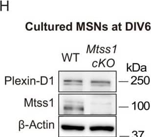
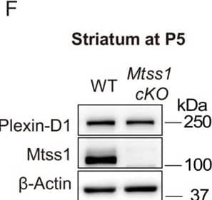
Detection of Plexin D1 by Western Blot The absence of Mtss1 does not affect medium spiny neuron (MSN) survival, dendritic arborization, and Plexin-D1 expression during striatonigral pathway development.(A) Immunohistochemistry staining for cleaved caspase 3 (CC3) in the striatum of wild-type (WT) or Mtss1 conditional knockout (cKO) mice. The white dotted boxes on the left images are shown in the inset images on the right at a better resolution. Scale bar, 25 μm. (B) Quantification of cell death by the number of CC3-positive cells in a 1 mm2 area covering the dorsal part of the striatum in WT or Mtss1 cKO mice. Error bars, mean ± SEM; ns p>0.05 by Mann‒Whitney test; WT, n = 20; Mtss1 cKO mice, n = 20 (five sections/mouse). (C) Representative images of Golgi staining at low (top panels) and high (bottom panels) magnification. Scale bars, 100 μm. Sholl analysis of dendritic morphology (D) and dendritic length (E) performed by using Neurolucida360 in 3D analysis. Error bars, mean ± SEM; ns p>0.05 by Student’s t-test; WT, n = 12, and Mtss1 cKO mice, n = 15, from three mice. (F, G) Western blot images and quantification of Plexin-D1 expression in the striatum of WT or Mtss1 cKO mice at P5. Error bars, mean ± SEM; *p<0.05 by Student’s t-test; WT mice, n = 3, and Mtss1 cKO mice, n = 4. (H) Plexin-D1 expression in MSNs obtained from the striatum of WT or Mtss1 cKO mice at P0 and measured at DIV6 in culture. (I) Quantification of the western blots shown in (H). Error bars, mean ± SEM; *p<0.05, by Student’s t-test; n = 3 for WT, n = 3 for KO in three independent experiments. Figure 7—figure supplement 3—source data 1.Raw uncropped western blot & gel images. Western blots shown in Figure 7—figure supplement 3F and H.Raw uncropped western blot & gel images. Western blots shown in Figure 7—figure supplement 3F and H. Image collected and cropped by CiteAb from the following open publication (https://elifesciences.org/articles/96891), licensed under a CC-BY license. Not internally tested by R&D Systems.
 View Larger
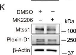
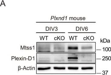
Detection of Plexin D1 by Western Blot In cultured medium spiny neurons (MSNs), Mtss1 expression is directly regulated by Sema3E-Plexin-D1 signaling through the AKT pathway.(A) WB images showing Mtss1 expression in MSNs derived from the striatum of wild-type (WT) or Plxnd1 conditional knockout (cKO) mice at P0 & measured at DIV3 & DIV6 in culture. (B) Quantification of band intensity in (A). Two-way ANOVA with Tukey’s post hoc correction for multiple comparisons; n = 3. (C) Mtss1 expression in MSNs obtained from the striatum of WT or Sema3e KO mice at P0 & measured at DIV6 in culture. (D) Quantification of the blots shown in (C). Student’s t-test; n = 5 for WT, n = 5 for KO in five independent experiments. (E) Schematic illustration of the experimental strategy for Sema3E-ligand or MK2206, an AKT inhibitor treatment in MSN culture. (F, G) WB images showing Mtss1 expression after AP-Sema3E (2 nM) treatment in cultured MSNs derived from Sema3e KO mice or Plxnd1 cKO mice. (H, I) Quantification of (F, G). Student’s t-test; AP, n = 4, AP-sema3E, n = 4 for sema3e KO mice, AP n = 4, AP-sema3E n = 4 for Plxnd1 cKO mice in three independent experiments. (J, K) WB to analyze the expression of Mtss1 & Plexin-D1 after MK2206 (100 nM) treatment in cultured MSNs & subsequent quantification for band intensity (L, M). Student’s t-test; n = 6 for sham, n = 6 for MK2206 in six independent experiments. (N O) WB image & analysis showing Mtss1 expression in Sema3e knockout MSNs treated with MK2206 after incubation with AP-Sema3E. Two-way ANOVA with Tukey’s post hoc correction for multiple comparisons; n = 5 in five independent experiments. Error bars, mean ± SEM; *p<0.05, **p<0.01, ***p<0.001 by indicated statistical tests. Figure 2—source data 1.WBs shown in Figure 2A, C, F, G, J, K, & N.WBs shown in Figure 2A, C, F, G, J, K, & N. Image collected & cropped by CiteAb from the following open publication (https://elifesciences.org/articles/96891), licensed under a CC-BY license. Not internally tested by R&D Systems.
 View Larger
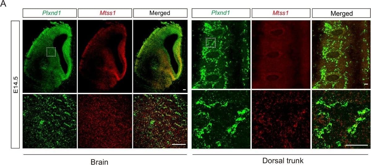
Detection of Mouse Plexin D1 by Immunohistochemistry No Mtss1 was found in endothelial cells at E14.5, and no vascular defects were observed in Mtss1-conditional knockout (KO) mice.(A) Fluorescence in situ hybridization for Plxnd1 mRNA (green) and Mtss1 mRNA (red) in the brain and dorsal trunk at E14.5. White dotted boxes are shown in the inset image on the bottom. Scale bars, 100 μm for brain, 50 μm for dorsal trunk. (B) 3D vascular reconstruction analysis images after CD31 immunostaining and tissue clearing obtained using multifunctional fast confocal microscopy Dragonfly 502w. (C) Western blotting to analyze Mtss1 expression after AP-Sema3E (2 nM) treatment in human umbilical vein endothelial cells (HUVECs) or human cortical microvessel endothelial cells (HCMEC/D3). Error bars, mean ± SEM; ns p>0.05 by Mann‒Whitney test; AP, n = 4, AP-Sema3E, n = 4 for HUVECs, ns p>0.05 by Student’s t-test; AP, n = 4, AP-Sema3E, n = 4 for HCMEC/D3s in four dependent experiments. Figure 8—figure supplement 1—source data 1.Western blots shown in Figure 8—figure supplement 1C.Western blots shown in Figure 8—figure supplement 1C. Image collected and cropped by CiteAb from the following open publication (https://elifesciences.org/articles/96891), licensed under a CC-BY license. Not internally tested by R&D Systems.
 View Larger
Detection of Plexin D1 by Western Blot In cultured medium spiny neurons (MSNs), Mtss1 expression is directly regulated by Sema3E-Plexin-D1 signaling through the AKT pathway.(A) WB images showing Mtss1 expression in MSNs derived from the striatum of wild-type (WT) or Plxnd1 conditional knockout (cKO) mice at P0 & measured at DIV3 & DIV6 in culture. (B) Quantification of band intensity in (A). Two-way ANOVA with Tukey’s post hoc correction for multiple comparisons; n = 3. (C) Mtss1 expression in MSNs obtained from the striatum of WT or Sema3e KO mice at P0 & measured at DIV6 in culture. (D) Quantification of the blots shown in (C). Student’s t-test; n = 5 for WT, n = 5 for KO in five independent experiments. (E) Schematic illustration of the experimental strategy for Sema3E-ligand or MK2206, an AKT inhibitor treatment in MSN culture. (F, G) WB images showing Mtss1 expression after AP-Sema3E (2 nM) treatment in cultured MSNs derived from Sema3e KO mice or Plxnd1 cKO mice. (H, I) Quantification of (F, G). Student’s t-test; AP, n = 4, AP-sema3E, n = 4 for sema3e KO mice, AP n = 4, AP-sema3E n = 4 for Plxnd1 cKO mice in three independent experiments. (J, K) WB to analyze the expression of Mtss1 & Plexin-D1 after MK2206 (100 nM) treatment in cultured MSNs & subsequent quantification for band intensity (L, M). Student’s t-test; n = 6 for sham, n = 6 for MK2206 in six independent experiments. (N O) WB image & analysis showing Mtss1 expression in Sema3e knockout MSNs treated with MK2206 after incubation with AP-Sema3E. Two-way ANOVA with Tukey’s post hoc correction for multiple comparisons; n = 5 in five independent experiments. Error bars, mean ± SEM; *p<0.05, **p<0.01, ***p<0.001 by indicated statistical tests. Figure 2—source data 1.WBs shown in Figure 2A, C, F, G, J, K, & N.WBs shown in Figure 2A, C, F, G, J, K, & N. Image collected & cropped by CiteAb from the following open publication (https://elifesciences.org/articles/96891), licensed under a CC-BY license. Not internally tested by R&D Systems.
 View Larger
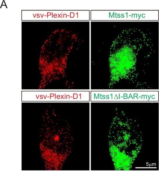
Detection of Mouse Plexin D1 by Immunohistochemistry No significant alteration in the expression of vsv-Plexin-D1 or Mtss1-myc or Mtss1 delta I-BAR-myc in the medium spiny neuron (MSN) soma.(A) Representative image of Immunocytochemistry for vsv-Plexin-D1 (red), Mtss1-myc or Mtss1 delta I-BAR -myc (green) in the cell body of cultured MSNs transfected with vsv-Plexin-D1 and Mtss1-myc or Mtss1 delta I-BAR-myc, using Mtss1-null mice as a background. (B) Quantification of the fluorescence intensity in the cell body of (A). Error bars, mean ± SEM; ns p>0.05 by two-way ANOVA with Tukey’s post hoc correction for multiple comparisons; vsv-Plexin-D1+Mtss1 myc, n = 5, and vsv-Plexin-D1+Mtss1 delta I-BAR-myc, n = 5. Scale bar, 5 μm. Image collected and cropped by CiteAb from the following open publication (https://elifesciences.org/articles/96891), licensed under a CC-BY license. Not internally tested by R&D Systems.
 View Larger
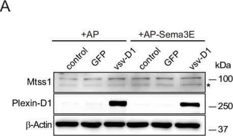
Detection of Plexin D1 by Western Blot Expression of Mtss1 induces I-BAR domain-dependent morphological changes in COS7 cells, generating protrusions.(A) Western blot images showing that weakly expression of endogenous Mtss1 was not altered by overexpression of Plexin-D1 with or without Sema3E in COS7 cells. Asterisk indicates nonspecific band. (B) Quantification of the band intensity in (A). Error bars, mean ± SEM; ns p>0.05 by two-way ANOVA with Bonferroni’s post hoc correction for multiple comparisons; n = 3. (C) Schematics describing the full-length construct of Mtss1-myc and its deletion mutant constructs (Mtss1 delta I-BAR-myc, Mtss1 delta WH2-myc, and I-BAR-myc). (D) Immunocytochemistry images taken after overexpression of each construct. Constructs show the I-BAR domain leading to diverse cell protrusion morphologies. Some of the protrusions were excessively spiked or thin and long (arrowheads). Overexpression of the I-BAR domain only (I-BAR-myc) can induce extreme protrusion structures. Scale bar, 20 μm.Figure 3—figure supplement 1—source data 1.Western blots shown in Figure 3—figure supplement 1A.Western blots shown in Figure 3—figure supplement 1A. Image collected and cropped by CiteAb from the following open publication (https://elifesciences.org/articles/96891), licensed under a CC-BY license. Not internally tested by R&D Systems.
 View Larger
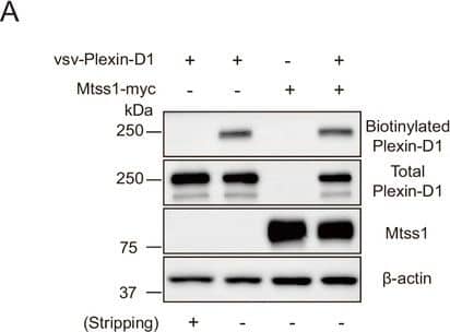
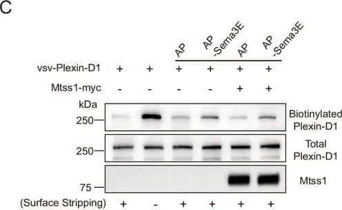
Detection of Plexin D1 by Western Blot Mtss1 expression alters Plexin-D1 localization to the protrusion structure in COS7 cells without affecting its endocytosis or Sema3E binding.(A) Cell surface biotinylation and subsequent Western blot analysis to analyze the surface localization of Plexin-D1 in COS7 cells. Image collected and cropped by CiteAb from the following open publication (https://elifesciences.org/articles/96891), licensed under a CC-BY license. Not internally tested by R&D Systems.
 View Larger
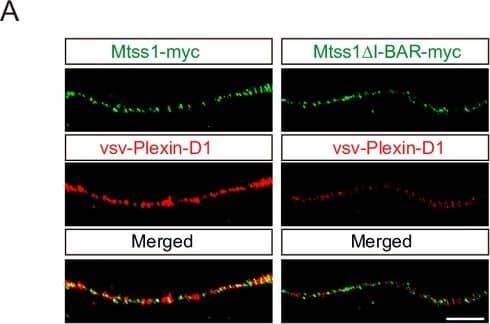
Detection of Mouse Plexin D1 by Immunohistochemistry Mtss1 facilitates Plexin-D1 transport to the growth cone in cultured Drd1a-positive medium spiny neurons (MSNs).(A) Immunocytochemistry for Mtss1-myc or Mtss1 delta I-BAR -myc (green), vsv-Plexin-D1 (red) in the axons of MSNs transfected with vsv-Plexin-D1 and Mtss1-myc or Mtss1 delta I-BAR-myc, using Mtss1-null mice as a background. The images were acquired using structured illumination microscopy (N-SIM). Scale bar, 5 μm. Image collected and cropped by CiteAb from the following open publication (https://elifesciences.org/articles/96891), licensed under a CC-BY license. Not internally tested by R&D Systems.
 View Larger
Detection of Plexin D1 by Western Blot Mtss1 facilitates Plexin-D1 transport to the growth cone in cultured Drd1a-positive medium spiny neurons (MSNs). Quantification of colocalization by Pearson’s correlation coefficient calculated using Costes’ randomized pixel scrambled image method. Student’s t-test; vsv-Plexin-D1+Mtss1-myc, n = 21, and vsv-Plexin-D1+Mtss1 delta IBAR-myc, n = 14. Image collected and cropped by CiteAb from the following open publication (https://elifesciences.org/articles/96891), licensed under a CC-BY license. Not internally tested by R&D Systems.
 View Larger
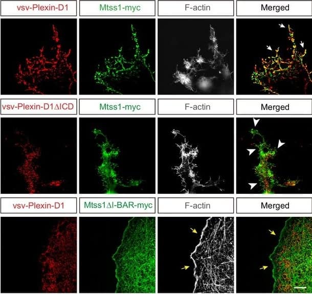
Detection of Mouse Plexin D1 by Immunohistochemistry Mtss1 expression alters Plexin-D1 localization to the protrusion structure in COS7 cells without affecting its endocytosis or Sema3E binding. (F) Immunocytochemistry for vsv-Plexin-D1 (red), Mtss1-myc (green), and F-actin (gray) in COS7 cells. Images were obtained by structured illumination microscopy (N-SIM). White arrows (top) indicate colocalized Plexin-D1 and Mtss1 in the protrusion structure. White arrowheads (middle) indicate high Mtss1 levels localized in cell protrusions without Plexin-D1. Yellow arrows (bottom) indicate the normal cell surface with Mtss1 delta I-BAR but no Plexin-D1 colocalization. Scale bar, 5 μm. Image collected and cropped by CiteAb from the following open publication (https://elifesciences.org/articles/96891), licensed under a CC-BY license. Not internally tested by R&D Systems.
 View Larger
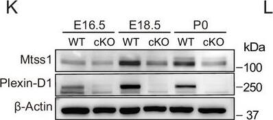
Detection of Mouse Plexin D1 by Western Blot Sema3E-Plexin-D1 signaling induces Mtss1 expression selectively in developing striatonigral projecting neurons. (K) Western blot images showing the expression of Mtss1 and Plexin-D1 in the striatum of WT or Plxnd1 cKO mice at different developmental stages ranging from embryonic day 16.5 (E16.5) to postnatal day 0 (P0). Image collected and cropped by CiteAb from the following open publication (https://elifesciences.org/articles/96891), licensed under a CC-BY license. Not internally tested by R&D Systems.
Reconstitution Calculator
Preparation and Storage
- 12 months from date of receipt, -20 to -70 °C as supplied.
- 1 month, 2 to 8 °C under sterile conditions after reconstitution.
- 6 months, -20 to -70 °C under sterile conditions after reconstitution.
Background: Plexin D1
Plexin D1 is a type I transmembrane glycoprotein that is the prototype of the plexin D subfamily of semaphorin receptors (1, 2). Human Plexin D1 contains a 46 amino acid (aa) signal sequence, a 1225 aa extracellular domain (ECD), a 21 aa transmembrane domain, and a 633 aa cytoplasmic domain that includes features common to other plexins (1). The human Plexin D1 ECD shares 89% identity with mouse Plexin D1, and ~84-92% aa identity based on incomplete sequences of rat, bovine, porcine and canine Plexin D1. It contains a sema domain, two plexin-semaphorin-integrin (PSI) or Met-related sequence (MRS) cysteine-rich motifs, and three glycine/proline-rich IPT/TIG domains which are immunoglobulin-like domains found in plexins, transcription factors, and the scatter factor receptors Met and Ron (1, 2). Isoforms of 1787 and 1747 aa have been sequenced; these contain a 178 aa N-terminal deletion with or without a longer alternate C-terminus (3). Like other Sema/plexin interactions, Plexin D1 interacts with Sema3C or Sema4A via neuropilins. Interaction with Sema3E, however, is direct (4). Plexin D1/Sema3E interaction mediates vascular guidance during development or angiogenesis; deletion of either molecule results in similar, profound cardiac abnormalities (4, 5). Plexin D1 is also expressed in lymphocytes, osteoblasts, the neural crest and the central nervous system during development (2, 6). In the brain, the presence of neuropilin can change Plexin D1/Sema3E interaction from an attractive to a repulsive signal (7, 8). Plexin D1 directs migration of thymocytes to the thymic medulla, probably through repulsion of Sema3E (9). Endothelial cell Plexin D1 binding to Sema4A can oppose VEGF and suppresses tumor angiogenesis, and expression of Sema3E correlates inversely with tumor metastasis, indicating that Plexin D1 is anti-metastatic in the presence of its ligands (10, 11).
- Negishi, M. et al. (2005) Cell. Mol. Life Sci. 62:1363.
- Van Der Zwaag, B. et al. (2002) Dev. Dyn. 225:336.
- Entrez protein Accession # Q9Y4D7, EAW79239, EAW79240.
- Gu, C. et al. (2005) Science 307:265.
- Gitler, A.D. et al. (2004) Developmental Cell 7:107.
- Zhang, Y. et al. (2009) Dev. Biol. 325:82.
- Chauvet, S. et al. (2007) Neuron 56:807.
- Pecho-Vrieseling, E. et al. (2009) Nature 459:842.
- Choi, Y.I. et al. (2008) Immunity 29:888.
- Toyofuku, T. et al. (2007) EMBO J. 26:1373.
- Roodink, I. et al. (2008) Am. J. Pathol. 173:1873.
Product Datasheets
Citations for Human Plexin D1 Antibody
R&D Systems personnel manually curate a database that contains references using R&D Systems products. The data collected includes not only links to publications in PubMed, but also provides information about sample types, species, and experimental conditions.
15
Citations: Showing 1 - 10
Filter your results:
Filter by:
-
SEMA6A drives GnRH neuron-dependent puberty onset by tuning median eminence vascular permeability
Authors: Lettieri, A;Oleari, R;van den Munkhof, MH;van Battum, EY;Verhagen, MG;Tacconi, C;Spreafico, M;Paganoni, AJJ;Azzarelli, R;Andre', V;Amoruso, F;Palazzolo, L;Eberini, I;Dunkel, L;Howard, SR;Fantin, A;Pasterkamp, RJ;Cariboni, A;
Nature communications
Species: Transgenic Mouse
Sample Types: Whole Tissue
Applications: Immunohistochemistry -
Insulin-like Growth Factor 1, Growth Hormone, and Anti-Müllerian Hormone Receptors Are Differentially Expressed during GnRH Neuron Development
Authors: Alyssa J. J. Paganoni, Rossella Cannarella, Roberto Oleari, Federica Amoruso, Renata Antal, Marco Ruzza et al.
International Journal of Molecular Sciences
-
Combined omic analyses reveal autism-linked NLGN3 gene as a key developmental regulator of GnRH neuron biology and disease
Authors: Roberto Oleari, Antonella Lettieri, Stefano Manzini, Alyssa Paganoni, Valentina André, Paolo Grazioli et al.
Disease Models & Mechanisms
Species: Mouse
Sample Types: Whole Tissue
Applications: Immunohistochemistry -
Motor neurons use push-pull signals to direct vascular remodeling critical for their connectivity
Authors: Luis F. Martins, Ilaria Brambilla, Alessia Motta, Stefano de Pretis, Ganesh Parameshwar Bhat, Aurora Badaloni et al.
Neuron
Species: Monkey, Mouse
Sample Types: Cell Lysates, Whole Cells, Embryo, Whole Tissue
Applications: Immunohistochemistry, Western Blot, Neutralization, Immunocytochemistry -
A Novel Loss-of-Function SEMA3E Mutation in a Patient with Severe Intellectual Disability and Cognitive Regression
Authors: AJJ Paganoni, F Amoruso, J Porta Pela, B Calleja-Pé, V Vezzoli, P Duminuco, A Caramello, R Oleari, A Fernández-, A Cariboni
International Journal of Molecular Sciences, 2022-05-18;23(10):.
Species: Human, Mouse, Primate - C. aethiops
Sample Types: Whole Cells, Whole Tissue
Applications: ICC, IHC -
Semaphorin3A/PlexinA3 association with the Scribble scaffold for cGMP increase is required for apical dendrite development
Authors: J Szczurkows, A Guo, J Martin, SI Lee, E Martinez, CT Chien, TA Khan, R Singh, D Dadson, TS Tran, S Pautot, M Shelly
Cell Reports, 2022-03-15;38(11):110483.
Species: Mouse, Rat
Sample Types: Whole Cells
Applications: ICC, IHC -
CRMP4-mediated fornix development involves Semaphorin-3E signaling pathway
Authors: Benoît Boulan, Charlotte Ravanello, Amandine Peyrel, Christophe Bosc, Christian Delphin, Florence Appaix et al.
eLife
-
Post-endocytic sorting of Plexin-D1 controls signal transduction and development of axonal and vascular circuits
Authors: K Burk, E Mire, A Bellon, M Hocine, J Guillot, F Moraes, Y Yoshida, M Simons, S Chauvet, F Mann
Nat Commun, 2017-02-22;8(0):14508.
Species: Mouse
Sample Types: Whole Cells
Applications: ICC -
Chemorepellent Semaphorin 3E Negatively Regulates Neutrophil Migration In Vitro and In Vivo
Authors: Hesam Movassagh
J. Immunol, 2016-12-02;0(0):.
Species: Human
Sample Types: Whole Cells
Applications: Flow Cytometry, ICC -
Semaphorin 3G Provides a Repulsive Guidance Cue to Lymphatic Endothelial Cells via Neuropilin-2/PlexinD1
Cell Rep, 2016-11-22;17(9):2299-2311.
Species: Human
Sample Types: Cell Lysates
Applications: Western Blot -
Infantile hemangioma-derived stem cells and endothelial cells are inhibited by class 3 semaphorins
Authors: Hironao Nakayama, Lan Huang, Ryan P. Kelly, Clara R.L. Oudenaarden, Adelle Dagher, Nicole A. Hofmann et al.
Biochemical and Biophysical Research Communications
-
Dysfunctional SEMA3E signaling underlies gonadotropin-releasing hormone neuron deficiency in Kallmann syndrome
Authors: Anna Cariboni, Valentina André, Sophie Chauvet, Daniele Cassatella, Kathryn Davidson, Alessia Caramello et al.
Journal of Clinical Investigation
-
An image-based RNAi screen identifies SH3BP1 as a key effector of Semaphorin 3E-PlexinD1 signaling.
Authors: Tata A, Stoppel D, Hong S, Ben-Zvi A, Xie T, Gu C
J Cell Biol, 2014-05-19;205(4):573-90.
Species: Human
Sample Types: Whole Cells
Applications: IHC -
Dual role for Islet-1 in promoting striatonigral and repressing striatopallidal genetic programs to specify striatonigral cell identity.
Authors: Lu, Kuan-Min, Evans, Sylvia M, Hirano, Shinji, Liu, Fu-Chin
Proc Natl Acad Sci U S A, 2013-12-18;111(1):E168-77.
Species: Mouse
Sample Types: Whole Tissue
Applications: IHC-P -
Sema3E-Plexin D1 signaling drives human cancer cell invasiveness and metastatic spreading in mice.
Authors: Casazza A, Finisguerra V, Capparuccia L
J. Clin. Invest., 2010-07-26;120(8):2684-98.
Species: Human
Sample Types: Cell Lysates
Applications: Western Blot
FAQs
No product specific FAQs exist for this product, however you may
View all Antibody FAQsReviews for Human Plexin D1 Antibody
There are currently no reviews for this product. Be the first to review Human Plexin D1 Antibody and earn rewards!
Have you used Human Plexin D1 Antibody?
Submit a review and receive an Amazon gift card.
$25/€18/£15/$25CAN/¥75 Yuan/¥2500 Yen for a review with an image
$10/€7/£6/$10 CAD/¥70 Yuan/¥1110 Yen for a review without an image
